You have no items in your shopping cart.
Page Not Found
Cart summary
Item 1 of 4
Item 1 of 4
CNOT8 Antibody
Catalog Number: orb1263820

| Catalog Number | orb1263820 |
|---|---|
| Category | Antibodies |
| Description | CNOT8 Antibody |
| Target | CNOT8 |
| Clonality | Polyclonal |
| Species/Host | Rabbit |
| Isotype | Rabbit Ig |
| Conjugation | Unconjugated |
| Reactivity | Human |
| Predicted Reactivity | Mouse |
| Form/Appearance | Liquid |
| Concentration | batch dependent |
| Buffer/Preservatives | Supplied in PBS with 0.09% (W/V) sodium azide. |
| Immunogen | This CNOT8 antibody is generated from rabbits immunized with a KLH conjugated synthetic peptide between 227-255 amino acids from the C-terminal region of human CNOT8. |
| UniProt ID | Q9UFF9 |
| MW | 34 kDa |
| Tested applications | FC, IF, IHC-P, WB |
| Application notes | For WB starting dilution is: 1:1000For IF starting dilution is: 1:10~50For IHC-P starting dilution is: 1:10~50For FACS starting dilution is: 1:10~50 |
| Antibody Type | Primary Antibody |
| Storage | Maintain refrigerated at 2-8°C for up to 2 weeks. For long term storage store at -20°C in small aliquots to prevent freeze-thaw cycles. |
| Alternative names | CCR4-NOT transcription complex subunit 8, CAF1-lik Read more... |
| Note | For research use only |
| NCBI | Q9UFF9 |

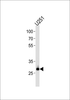
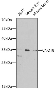
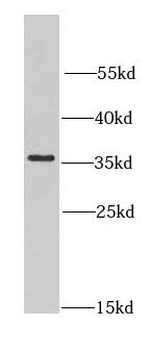
Western blot analysis in U251 cell line lysates (35 ug/lane).
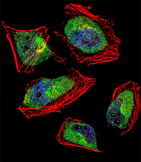
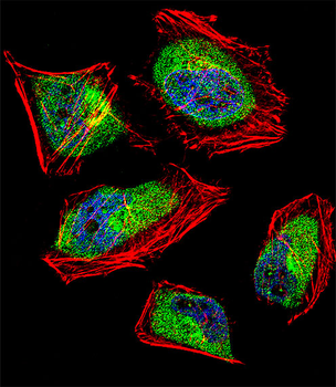
Fluorescent confocal image of Hela cell stained with CNOT8 Antibody.Hela cells were fixed with 4% PFA (20 min), permeabilized with Triton X-100 (0.1%, 10 min), then incubated with CNOT8 primary antibody (1:25). For secondary antibody, Alexa Fluor 488 conjugated donkey anti-rabbit antibody (green) was used (1:400).Cytoplasmic actin was counterstained with Alexa Fluor 555 (red) conjugated Phalloidin (7 units/ml). Nuclei were counterstained with DAPI (blue) (10 ug/ml, 10 min). CNOT8 immunoreactivity is localized to Cytoplasm significantly and Nucleus weakly.

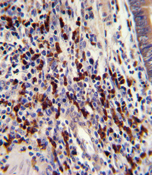
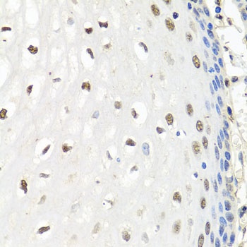
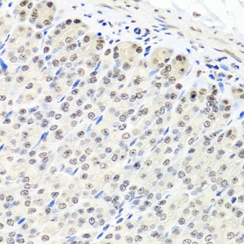
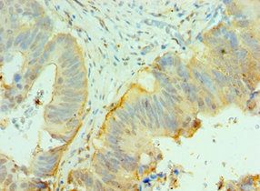
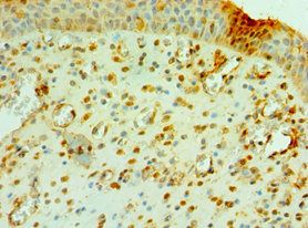
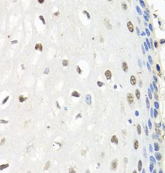
Formalin-fixed and paraffin-embedded human colon carcinoma with CNOT8 Antibody, which was peroxidase-conjugated to the secondary antibody, followed by DAB staining.
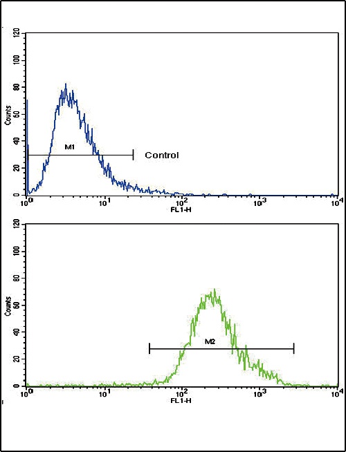
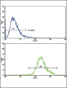
Flow cytometric analysis of CEM cells using CNOT8 Antibody (bottom histogram) compared to a negative control cell (top histogram). FITC-conjugated goat-anti-rabbit secondary antibodies were used for the analysis.
- Item 1 of 4
CNOT8 Antibody (C-term) [orb1930019]
FC, IF, IHC-P, WB
Mouse
Human
Rabbit
Polyclonal
Unconjugated
100 μl, 50 μl - Item 1 of 3
- Item 1 of 2
- Item 1 of 2
CNOT8 Antibody [orb625904]
ELISA, IHC, WB
Human, Mouse, Rat
Rabbit
Polyclonal
Unconjugated
100 μg, 50 μg - Item 1 of 1