You have no items in your shopping cart.
Description
Research Area
Cell Biology
Images & Validation
−Item 1 of 3
| Tested Applications | IF, IHC-P, WB |
|---|---|
| Dilution Range | IF - 1:10-50, WB - 1:1000, IHC-P-Leica - 1:10-50 |
| Reactivity | Human |
| Predicted Reactivity | Mouse, Rat |
Key Properties
−| Antibody Type | Primary Antibody |
|---|---|
| Host | Rabbit |
| Clonality | Polyclonal |
| Isotype | Rabbit IgG |
| Immunogen | This CLC4 antibody is generated from rabbits immunized with a KLH conjugated synthetic peptide between 663-689 amino acids from the C-terminal region of human CLC4. Antigen Region: 663-689 aa. |
| Target | CLCN4 |
| Molecular Weight | 84917 Da |
| Conjugation | Unconjugated |
Storage & Handling
−| Storage | Maintain refrigerated at 2-8°C for up to 2 weeks. For long term storage store at -20°C in small aliquots to prevent freeze-thaw cycles |
|---|---|
| Form/Appearance | Purified polyclonal antibody supplied in PBS with 0.09% (W/V) sodium azide. This antibody is prepared by Saturated Ammonium Sulfate (SAS) precipitation followed by dialysis against PBS. |
| Expiration Date | 12 months from date of receipt. |
| Disclaimer | For research use only |
Alternative Names
−H(+)/Cl(-) exchange transporter 4, Chloride channel protein 4, ClC-4, Chloride transporter ClC-4, CLCN4
Quality Guarantee
Explore bioreagents carefree to elevate your research. All our products are rigorously tested for performance. If a product does not perform as described on its datasheet, our scientific support team will provide expert troubleshooting, a prompt replacement, or a refund. For full details, please see our Terms & Conditions and Buying Guide. Contact us at [email protected].
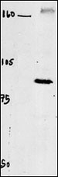
Western blot of chicken brain tissue incubated with CLC4 Antibody (C-term).
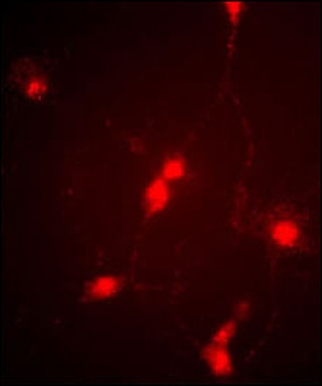
Immunofluorescence image of cultured chick retinal amacrine (neuronal) cells labeled with CLC4 Antibody (C-term).
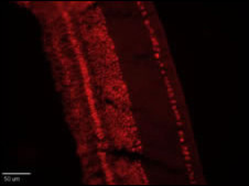
Retinae were collected from adult White Leghorn chicken and fixed in the eyecup for two hours in 4% Paraformaldehyde (in PBS). Retinae were then removed from the eyecups and incubated in 30% sucrose (in PBS) overnight. Retinal tissue was embedded and frozen in OCT compound and cut into ~15um sections on a cryotome. Sections were blocked in 5% normal goat serum (in 1% BSA/.1% saponin PBS) for one hour and then incubated at RT with 1:250 (1% BSA/.1% saponin PBS) ClC4 antibody for 1 hour. Sections were then washed in PBS (3X10minutes) and then treated with secondary antibody (1:500 Cy3) for one hour. After another PBS wash series, sections were coverslipped and antibody labeling was visualized at 20X with a Leica upright microscope using a TRITC filter set and Xenon lamp illumination. (Crousillac et al, 2003)
Quick Database Links
UniProt Details
− No UniProt data available
NCBI Reference Sequences
−Associated Accession Numbers
Curated reference sequences for the gene transcript and protein product| Protein | NP_001821.2 |
|---|
Documents Download
Datasheet
Product Information
Request a Document
Protocol Information
WB
Western Blot (IB, immunoblot)
IHC-P
Immunohistochemistry Paraffin
IF
Immunofluorescence
CLC4 Antibody (C-term) (orb1930081)

- 0.0
Based on 0 reviews
Participating in our Biorbyt product reviews program enables you to support fellow scientists by sharing your firsthand experience with our products.
Login to Submit a ReviewAvailable Sizes
Select a size below
Choose Conjugation or Carrier Free Version
Free Secondary Antibody (20 ul)0/0
Please add an antibody product to your cart first.