You have no items in your shopping cart.
Cart summary
Item 1 of 6
Item 1 of 6
CF150 Antibody
Catalog Number: orb1270774

| Catalog Number | orb1270774 |
|---|---|
| Category | Antibodies |
| Description | CF150 Antibody |
| Target | MB21D1 |
| Clonality | Polyclonal |
| Isotype | Rabbit Ig |
| Conjugation | Unconjugated |
| Reactivity | Human |
| Form/Appearance | Liquid |
| Concentration | batch dependent |
| Buffer/Preservatives | Supplied in PBS with 0.09% (W/V) sodium azide. |
| Immunogen | This CF150 antibody is generated from rabbits immunized with a KLH conjugated synthetic peptide between 266-295 amino acids from the Central region of human CF150. |
| UniProt ID | Q8N884 |
| MW | 59 kDa |
| Tested applications | FC, WB |
| Application notes | For FACS starting dilution is: 1:25For WB starting dilution is: 1:500-1:1000 |
| Antibody Type | Primary Antibody |
| Storage | Maintain refrigerated at 2-8°C for up to 2 weeks. For long term storage store at -20°C in small aliquots to prevent freeze-thaw cycles. |
| Alternative names | Cyclic GMP-AMP synthase, cGAMP synthase, cGAS, h-c Read more... |
| Note | For research use only |
| NCBI | Q8N884 |

Overlay histogram showing U-2OS cells stained with Antibody (green line). The cells were fixed with 2% paraformaldehyde (10 min) and then permeabilized with 90% methanol for 10 min. The cells were then icubated in 2% bovine serum albumin to block non-specific protein-protein interactions followed by the antibody (1:25 dilution) for 60 min at 37oC. The secondary antibody used was Goat-Anti-Rabbit IgG, DyLight 488 Conjugated Highly Cross-Adsorbed (NA168821) at 1/400 dilution for 40 min at 37oC. Isotype control antibody (blue line) was rabbit IgG (1 ug/1x10^6 cells) used under the same conditions. Acquisition of >10000 events was performed.
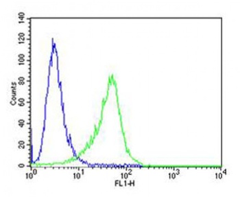
Flow cytometric analysis of A549 cells using CF150 Antibody (green) compared to an isotype control of rabbit IgG (blue). Antibody was diluted at 1:25 dilution. An Alexa Fluor 488 goat anti-rabbit lgG at 1:400 dilution was used as the secondary antibody.

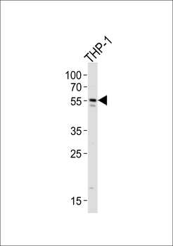
Western blot analysis of lysate from THP-1 cell line, using CF150 Antibody at 1:1000.
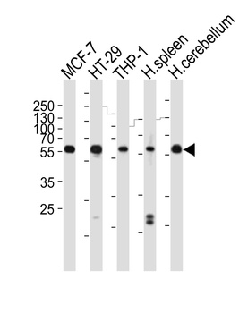
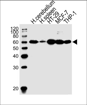
Western blot analysis of lysates from MCF-7, HT-29, THP-1 cell line, human spleen, human cerebellum tissue lysate (from left to right), using CF150 Antibody at 1:1000 at each lane.

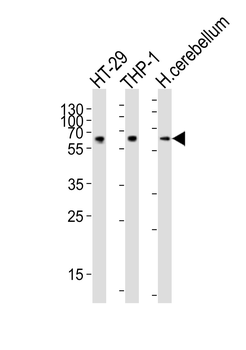
Western blot analysis of lysates from HT-29, THP-1 cell line, human cerebellum tissue lysate (from left to right), using CF150 Antibody at 1:1000 at each lane.

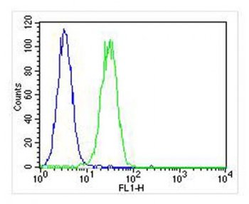
Flow cytometric analysis of MDA-MB231 cells (right histogram) compared to a negative control cell (left histogram). FITC-conjugated goat-anti-rabbit secondary antibodies were used for the analysis.
- Item 1 of 4
- Item 1 of 2