You have no items in your shopping cart.
Cart summary
Item 1 of 3
Item 1 of 3
CEA Antibody / Carcinoembryonic Antigen
Catalog Number: orb606358

| Catalog Number | orb606358 |
|---|---|
| Category | Antibodies |
| Description | This antibody recognizes proteins of 80-200kDa, identified as different members of CEA family. CEA is synthesized during development in the fetal gut and is re-expressed in increased amounts in intestinal carcinomas and several other tumors. This mAb reacts with nonspecific cross-reacting antigen (NCA). It shows no reaction with a variety of normal tissues and is suitable for staining of formalin/paraffin tissues. CEA is not found in benign glands, stroma, or malignant prostatic cells. Antibody to CEA is useful in detecting early foci of gastric carcinoma and in distinguishing pulmonary adenocarcinomas (60-70% are CEA+) from pleural mesotheliomas (rarely or weakly CEA+). Anti-CEA positivity is seen in adenocarcinomas from the lung, colon, stomach, esophagus, pancreas, gallbadder, urachus, salivary gland, ovary, and endocervix. |
| Clonality | Monoclonal |
| Species/Host | Mouse |
| Isotype | Mouse IgG1 |
| Conjugation | Unconjugated |
| Reactivity | Human |
| Immunogen | Purified full length human protein was used as the immunogen for the CEA antibody. |
| UniProt ID | P06731 |
| Tested applications | ELISA, IHC-P |
| Dilution range | ELISA (order BSA/sodium azide-free format for coating),Immunohistochemistry (FFPE): 1-2ug/ml for 30 min at RT (1) (2) |
| Application notes | Optimal dilution of the CEA antibody to be determined by the researcher. |
| Antibody Type | Primary Antibody |
| Clone Number | C66/1291 |
| Formula | 0.2 mg/ml in 1X PBS with 0.1 mg/ml BSA (US sourced) and 0.05% sodium azide |
| Storage | Maintain refrigerated at 2-8°C for up to 2 weeks. For long term storage store at -20°C in small aliquots to prevent freeze-thaw cycles. |
| Hazard Information | This CEA antibody is available for research use only. |
| Note | For research use only |
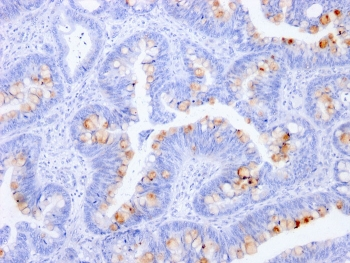
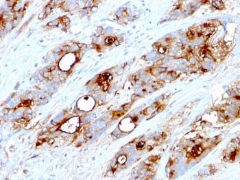
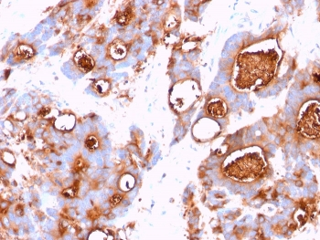
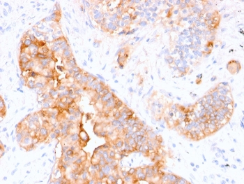
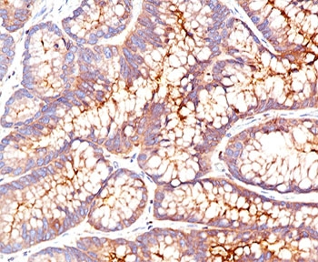
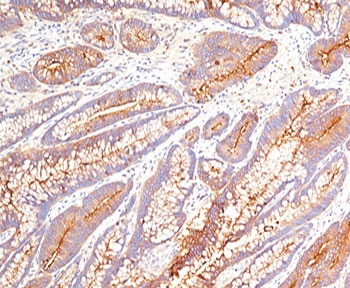
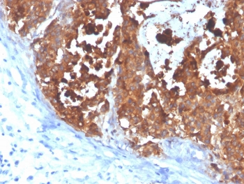
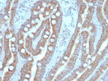
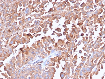
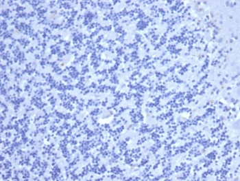
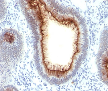
IHC staining of FFPE human colon with CEA antibody (clone C66/1291). HIER: boil tissue sections in pH9 10mM Tris with 1mM EDTA for 10-20 min and allow to cool before testing.
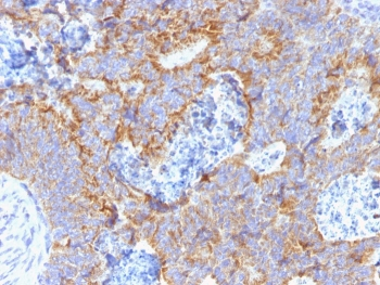
IHC staining of FFPE human colon with CEA antibody (clone C66/1291). HIER: boil tissue sections in pH9 10mM Tris with 1mM EDTA for 10-20 min and allow to cool before testing.
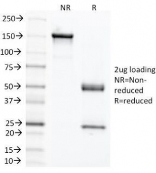
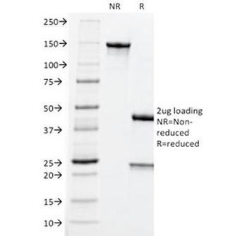
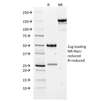
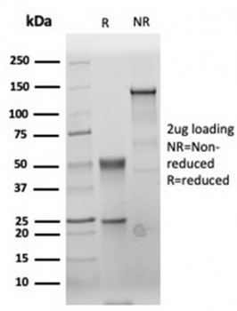
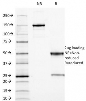
SDS-PAGE analysis of purified, BSA-free CEA antibody (clone C66/1291) as confirmation of integrity and purity.
- Item 1 of 5
CEA Antibody / Carcinoembryonic Antigen [orb248299]
IHC-P
Human
Mouse
Monoclonal
Unconjugated
20 μg, 100 μg - Item 1 of 3
CEA Antibody / Carcinoembryonic Antigen [orb248295]
IHC-P, WB
Human
Mouse
Monoclonal
Unconjugated
20 μg, 100 μg - Item 1 of 3
CEA Antibody / Carcinoembryonic Antigen [orb699698]
IHC-P
Human
Mouse
Monoclonal
Unconjugated
20 μg, 100 μg - Item 1 of 3
CEA Antibody / Carcinoembryonic Antigen [orb1151525]
IHC-P
Human
Rabbit
Recombinant
Unconjugated
20 μg, 100 μg - Item 1 of 2
CEA Antibody / Carcinoembryonic Antigen [orb385664]
IHC-P
Human
Mouse
Monoclonal
Unconjugated
20 μg, 100 μg