You have no items in your shopping cart.
Cart summary
Item 1 of 5
Item 1 of 5
CD59 Antibody (Center)
Catalog Number: orb1424640

| Catalog Number | orb1424640 |
|---|---|
| Category | Antibodies |
| Description | Rabbit polyclonal antibody to CD59. |
| Target | CD59 |
| Clonality | Polyclonal |
| Species/Host | Rabbit |
| Isotype | Rabbit IgG |
| Conjugation | Unconjugated |
| Reactivity | Human |
| Form/Appearance | Purified polyclonal antibody supplied in PBS with 0.09% (W/V) sodium azide. This antibody is purified through a protein A column, followed by peptide affinity purification. |
| Immunogen | Synthesized Peptide |
| UniProt ID | P13987 |
| MW | 14177 |
| Tested applications | FC, IF, IHC-P, WB |
| Dilution range | IF: 1:25, WB: 1:2000, IHC-P-Leica: 1:1000, IHC: 1:250, FC: 1:25 |
| Antibody Type | Primary Antibody |
| Clone Number | RB56691 |
| Storage | Maintain refrigerated at 2-8°C for up to 2 weeks. For long term storage store at -20°C in small aliquots to prevent freeze-thaw cycles |
| Alternative names | CD59 glycoprotein, 1F5 antigen, 20 kDa homologous Read more... |
| Note | For research use only |
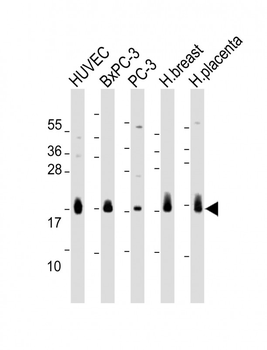
All lanes: Anti-CD59 Antibody (Center) at 1:2000 dilution. Lane 1: HUVEC whole cell lysate. Lane 2: BxPC-3 whole cell lysate. Lane 3: PC-3 whole cell lysate. Lane 4: Human breast lysate. Lane 5: Human placenta lysate.Lysates/proteins at 20 µg per lane. Secondary Goat Anti-Rabbit IgG, (H+L), Peroxidase conjugated at 1/10000 dilution. Predicted band size: 14 kDa. Blocking/Dilution buffer: 5% NFDM/TBST.
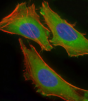
Immunofluorescent analysis of 4% paraformaldehyde-fixed, 0.1% Triton X-100 permeabilized HeLa (human cervical epithelial adenocarcinoma cell line) cells labeling CD59 at 1/25 dilution, followed by Dylight 488-conjugated goat anti-rabbit IgG secondary antibody at 1/200 dilution (green). Immunofluorescence image showing cytoplasm staining on HeLa cell line. Cytoplasmic actin is detected with Dylight 554 Phalloidin at 1/100 dilution (red). The nuclear counter stain is DAPI (blue).

Immunohistochemical analysis of paraffin-embedded human placent tissue performed on the Leica BOND RXm. Tissue was fixed with formaldehyde at room temperature; antigen retrieval was by heat mediation with a EDTA buffer (pH9.0). Samples were incubated with primary antibody for 1 hours at room temperature. A undiluted biotinylated CRF Anti-Polyvalent HRP Polymer antibody was used as the secondary antibody.
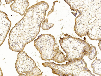
Immunohistochemical analysis of paraffin-embedded Human placenta section using Pink1. Diluted at 1:250 dilution. A undiluted biotinylated goat polyvalent antibody was used as the secondary, followed by DAB staining.
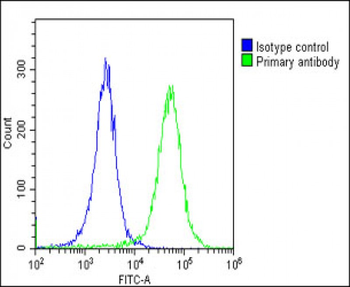
Overlay histogram showing HeLa cells stained (green line). The cells were fixed with 2% paraformaldehyde (10 min) and then permeabilized with 90% methanol for 10 min. The cells were then icubated in 2% bovine serum albumin to block non-specific protein-protein interactions followed by the antibody (1:25 dilution) for 60 min at 37°C. The secondary antibody used was Goat-Anti-Rabbit IgG, DyLight 488 Conjugated Highly Cross-Adsorbed at 1/200 dilution for 40 min at 37°C. Isotype control antibody (blue line) was rabbit IgG1 (1μg/1x10^6 cells) used under the same conditions. Acquisition of >10, 000 events was performed.
