You have no items in your shopping cart.
Cart summary
Item 1 of 15
Item 1 of 15
beta Catenin/CTNNB1 Antibody
Catalog Number: orb402308
| Catalog Number | orb402308 |
|---|---|
| Category | Antibodies |
| Description | beta Catenin/CTNNB1 Antibody |
| Species/Host | Rabbit |
| Clonality | Polyclonal |
| Tested applications | ELISA, FC, ICC, IF, IHC, WB |
| Reactivity | Human, Mouse, Rat |
| Isotype | Rabbit IgG |
| Immunogen | E. coli-derived human beta Catenin recombinant protein (Position: A2-K233). |
| Concentration | Adding 0.2 ml of distilled water will yield a concentration of 500 μg/ml. |
| Dilution range | Western blot, 0.1-0.5μg/ml Immunohistochemistry (Paraffin-embedded Section), 0.5-1μg/ml Immunocytochemistry/Immunofluorescence, 2μg/ml Immunofluorescence, 5μg/ml Flow Cytometry, 1-3μg/1x106 cellsbr> Direct ELISA, 0.1-0.5μg/ml |
| Form/Appearance | Lyophilized |
| Conjugation | Unconjugated |
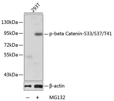
| MW | 95 kDa |
| UniProt ID | P35222 |
| Storage | Store at -20˚C for one year from date of receipt. After reconstitution, at 4˚C for one month. It can also be aliquotted and stored frozen at -20˚C for six months. Avoid repeated freeze-thaw cycles. |
| Alternative names | Catenin beta-1; Beta-catenin; CTNNB1; CTNNB; OK/SW Read more... |
| Note | For research use only |
| Application notes | Tested Species: In-house tested species with positive results. By Heat: Boiling the paraffin sections in 10mM citrate buffer, pH6.0, for 20mins is required for the staining of formalin/paraffin sections. Other applications have not been tested. Optimal dilutions should be determined by end users. . Add 0.2ml of distilled water will yield a concentration of 500ug/ml. |
| Expiration Date | 12 months from date of receipt. |

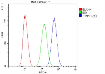
Flow Cytometry analysis of A549 cells using anti-CTNNB1 antibody (Blue line).Isotype control antibody (Green line) was rabbit IgG .Unlabelled sample (Red line) was also used as a control.
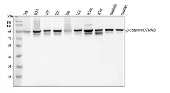
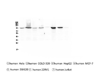
WB analysis using anti-CTNNB1 antibody.Lane 1:human Hela cell; 2:human MCF-7 cell; 3:human A431 cell; 4:human RT4 cell; 5:human SiHa cell; 6:human T47D cell; 7:rat brain tissue; 8:rat liver tissue; 9:mouse brain tissue; 10:mouse liver tissue.
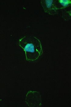
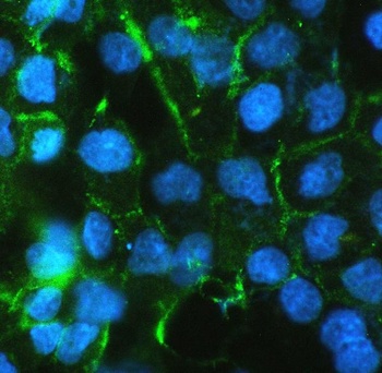
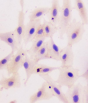
IF analysis of CTNNB1 using anti-CTNNB1 antibody.CTNNB1 was detected in immunocytochemical section of A431 cell.
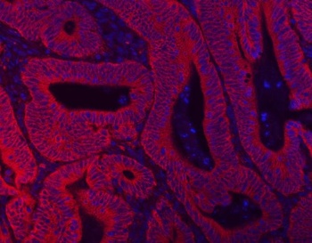
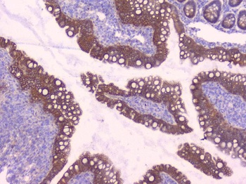
IF analysis of CTNNB1 using anti-CTNNB1 antibody. CTNNB1 was detected in a paraffin-embedded section of human intestine cancer tissue.
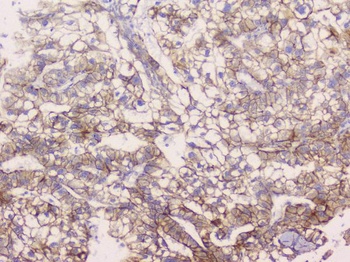
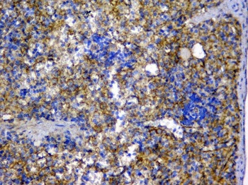
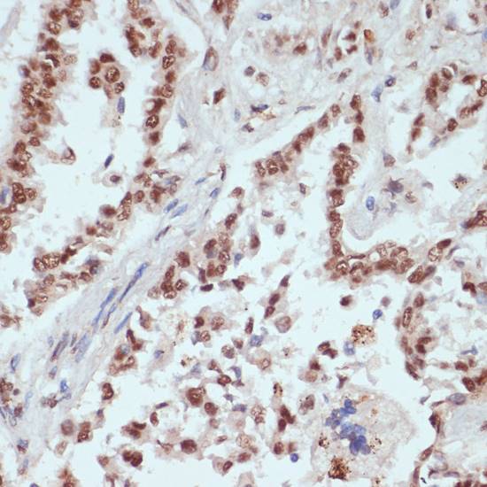
IHC analysis of CTNNB1 using anti-CTNNB1 antibody.CTNNB1 was detected in paraffin-embedded section of human liver cancer tissue.
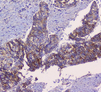
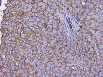
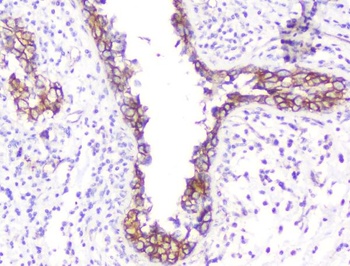
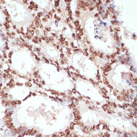
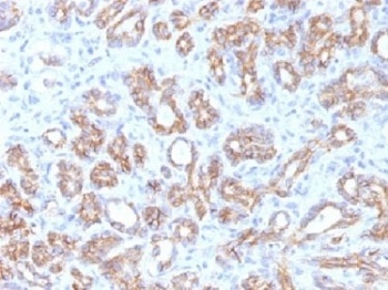
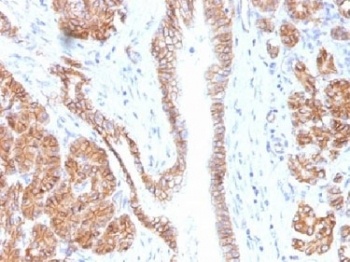
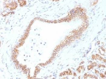
IHC analysis of CTNNB1 using anti-CTNNB1 antibody.CTNNB1 was detected in paraffin-embedded section of human prostatic cancer tissue.
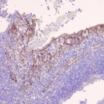
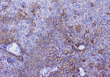
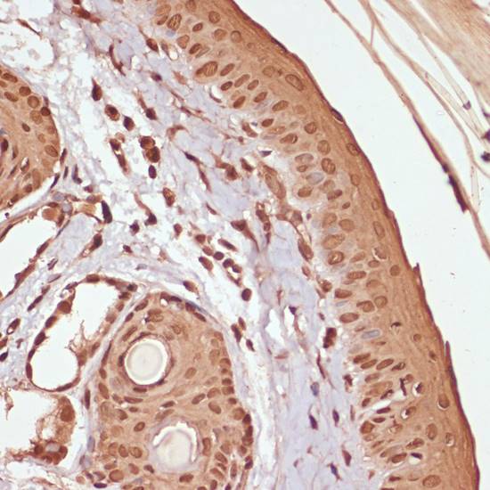
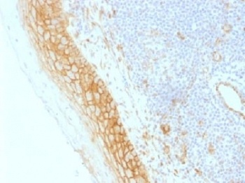
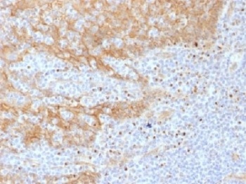
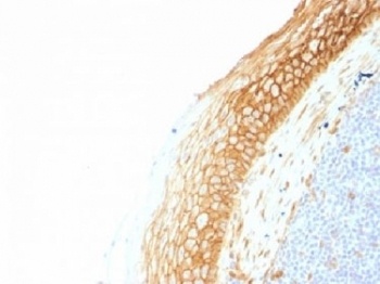
IHC analysis of CTNNB1 using anti-CTNNB1 antibody.CTNNB1 was detected in paraffin-embedded section of human tonsil tissue.
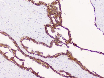
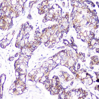
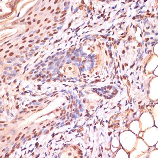
IHC analysis of CTNNB1 using anti-CTNNB1 antibody.CTNNB1 was detected in paraffin-embedded section of human mammary cancer tissue.
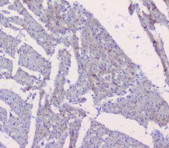
IHC analysis of CTNNB1 using anti-CTNNB1 antibody.CTNNB1 was detected in paraffin-embedded section of mouse heart tissue.
IHC analysis of CTNNB1 using anti-CTNNB1 antibody.CTNNB1 was detected in paraffin-embedded section of mouse liver tissue.
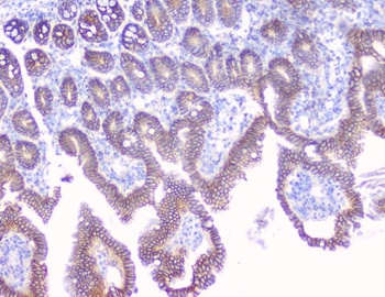
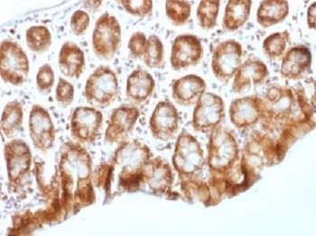
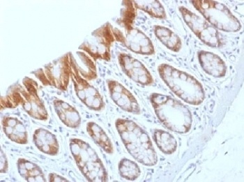
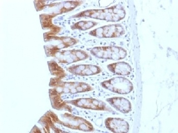
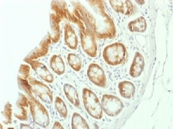
IHC analysis of CTNNB1 using anti-CTNNB1 antibody.CTNNB1 was detected in paraffin-embedded section of mouse intestine tissue.
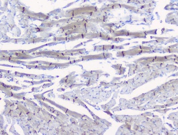
IHC analysis of CTNNB1 using anti-CTNNB1 antibody.CTNNB1 was detected in paraffin-embedded section of rat heart tissue.
IHC analysis of CTNNB1 using anti-CTNNB1 antibody.CTNNB1 was detected in paraffin-embedded section of rat liver tissue.
IHC analysis of CTNNB1 using anti-CTNNB1 antibody.CTNNB1 was detected in paraffin-embedded section of rat spleen tissue.
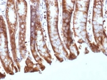
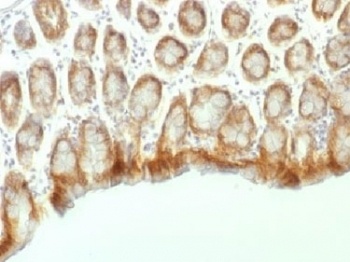
IHC analysis of CTNNB1 using anti-CTNNB1 antibody.CTNNB1 was detected in paraffin-embedded section of rat intestine tissue.
- Item 1 of 6
beta Catenin CTNNB1 Antibody (monoclonal, 1F6) [orb421109]
FC, ICC, IF, IHC, WB
Human, Mouse, Rat
Mouse
Monoclonal
Unconjugated
10 μg, 100 μg - Item 1 of 5
CTNNB1 (Phospho-S33/S37/T41) antibody [orb536885]
IHC
Human, Mouse, Rat
Unconjugated
200 μl, 100 μl, 50 μl - Item 1 of 4
- Item 1 of 4
- Item 1 of 4
Submit a review
Filter by Rating

- 5 stars

- 4 stars
- 3 stars
- 2 stars
- 1 stars