You have no items in your shopping cart.
Description
Research Area
Disease Biomarkers, Neuroscience
Images & Validation
−Item 1 of 2
| Tested Applications | IHC, WB |
|---|---|
| Dilution Range | WB (1:1000), IHC (1:400) |
| Reactivity | Human, Mouse, Rat |
| Application Notes |
Key Properties
−| Host | Rabbit |
|---|---|
| Clonality | Polyclonal |
| Immunogen | NM_003458.3 (AA 786-1041) N-terminal his-tagged fusion protein |
| Target | Bassoon |
| Molecular Weight | 420kDa |
| Purification | Protein A Purified |
| Conjugation | PerCP |
Storage & Handling
−| Storage | Conjugated antibodies should be stored according to the product label |
|---|---|
| Buffer/Preservatives | 95.46mM Phosphate, 2.48mM MES and 2mM EDTA |
| Concentration | 1 mg/ml |
| Expiration Date | 12 months from date of receipt. |
| Disclaimer | For research use only |
Alternative Names
−Bassoon, BSN, ZNF231, Zinc finger protein 231, Neuronal double zinc finger protein, KIAA0434
Similar Products
−- Item 1 of 2
- Item 1 of 2

Bassoon Rabbit pAb, PerCP-Cy7 conjugated [orb2852405]
IF
Human, Rat
Mouse
Rabbit
Polyclonal
PerCP/Cy7
100 μlBassoon Rabbit Polyclonal Antibody (PerCP-Cy5.5) [orb2559034]
IF
Human, Rat
Mouse
Rabbit
Polyclonal
PerCP/Cy5.5
100 μlBassoon Rabbit Polyclonal Antibody (PerCP) [orb2559035]
IF
Human, Rat
Mouse
Rabbit
Polyclonal
PerCP
100 μl
Quality Guarantee
Explore bioreagents carefree to elevate your research. All our products are rigorously tested for performance. If a product does not perform as described on its datasheet, our scientific support team will provide expert troubleshooting, a prompt replacement, or a refund. For full details, please see our Terms & Conditions and Buying Guide. Contact us at [email protected].
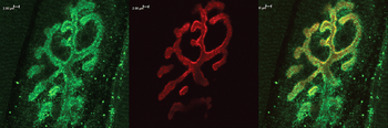
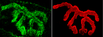
Immunohistochemistry analysis using Rabbit Anti-Bassoon Polyclonal Antibody. Tissue: Muscle. Species: Mouse. Primary Antibody: Rabbit Anti-Bassoon Polyclonal Antibody at 1:400. Secondary Antibody: Alexa Fluor 488 Goat Anti-Rabbit. Counterstain: BTX (red). Localization: Selective staining of the NMJ.
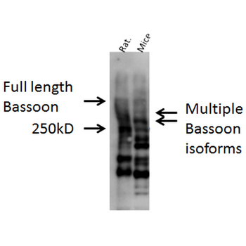
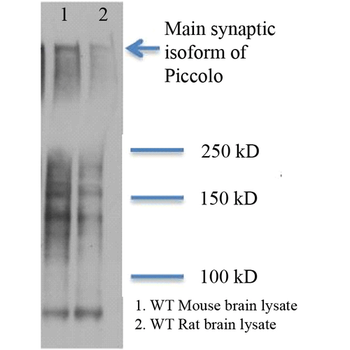
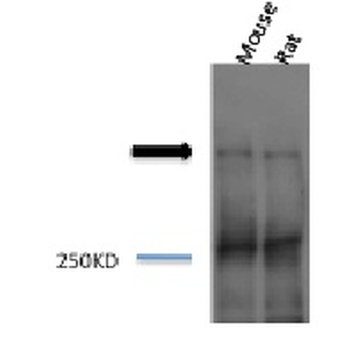
Western blot analysis of Mouse, Rat brain cell lysates showing detection of Bassoon protein using Rabbit Anti-Bassoon Polyclonal Antibody. Primary Antibody: Rabbit Anti-Bassoon Polyclonal Antibody at 1:1000.
Quick Database Links
UniProt Details
− No UniProt data available
NCBI Gene Details
− No NCBI Gene data available
NCBI Reference Sequences
−Associated Accession Numbers
Curated reference sequences for the gene transcript and protein product| Protein | NP_003449.2 |
|---|
Documents Download
Datasheet
Product Information
Request a Document
Protocol Information
WB
Western Blot (IB, immunoblot)
IHC
Immunohistochemistry
Bassoon Antibody (PerCP) (orb152189)

- 0.0
Based on 0 reviews
Participating in our Biorbyt product reviews program enables you to support fellow scientists by sharing your firsthand experience with our products.
Login to Submit a ReviewAvailable Sizes
Select a size below
Choose Conjugation or Carrier Free Version
Free Secondary Antibody (20 ul)0/0
Please add an antibody product to your cart first.