You have no items in your shopping cart.
Page Not Found
Cart summary
Item 1 of 4
Item 1 of 4
ATG4D Antibody (C-term)
Catalog Number: orb1933457

| Catalog Number | orb1933457 |
|---|---|
| Category | Antibodies |
| Description | Purified Rabbit Polyclonal Antibody (Pab) |
| Target | This ATG4D antibody is generated from rabbits immunized with a KLH conjugated synthetic peptide between 441-470 amino acids from the C-terminal region of human ATG4D. |
| Clonality | Polyclonal |
| Species/Host | Rabbit |
| Isotype | Rabbit IgG |
| Conjugation | Unconjugated |
| Reactivity | Human |
| Form/Appearance | Purified polyclonal antibody supplied in PBS with 0.09% (W/V) sodium azide. This antibody is prepared by Saturated Ammonium Sulfate (SAS) precipitation followed by dialysis against PBS. |
| UniProt ID | Q86TL0 |
| MW | 52922 Da |
| Tested applications | IF, IHC-P, WB |
| Dilution range | IF: 1:100, WB: 1:1000, WB: 1:1000, IHC-P: 1:50~100 |
| Antibody Type | Primary Antibody |
| Clone Number | RB7567 |
| Storage | Maintain refrigerated at 2-8°C for up to 2 weeks. For long term storage store at -20°C in small aliquots to prevent freeze-thaw cycles |
| Alternative names | Cysteine protease ATG4D, 3422-, AUT-like 4 cystein Read more... |
| Note | For research use only |
| NCBI | NP_116274.3, NP_001268433.1 |
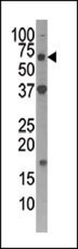
Western blot analysis of APG4D Pab in Y79 cell lysate.
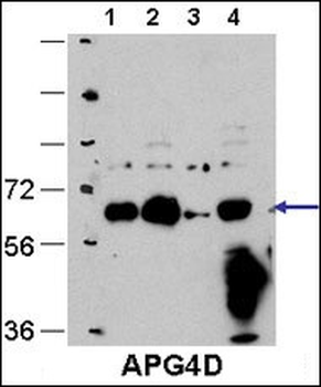
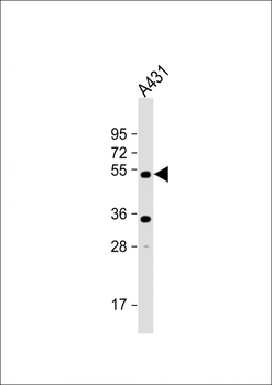
Cos7, HEK293, MEF, and Hela cells, left to right respectively.
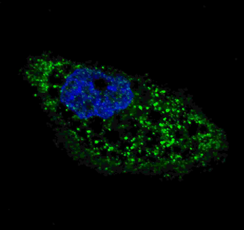
Fluorescent image of U251 cells stained with ATG4D (C-term) antibody. U251 cells were treated with Chloroquine (50 μM, 16 h), then fixed with 4% PFA (20 min), permeabilized with Triton X-100 (0.2%, 30 min). Cells were then incubated with ATG4D (C-term) primary antibody (1:100, 2 h at room temperature). For secondary antibody, Alexa Fluor 488 conjugated donkey anti-rabbit antibody (green) was used (1:1000, 1 h). Nuclei were counterstained with Hoechst 33342 (blue) (10 μg/ml, 5 min). ATG4D immunoreactivity is localized to autophagic vacuoles in the cytoplasm of U251 cells.
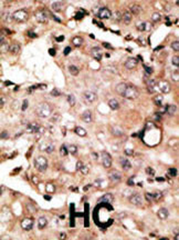
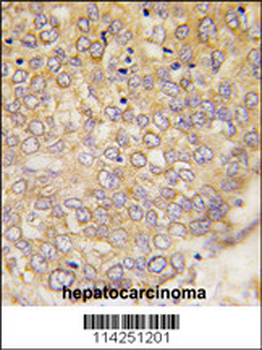
Formalin-fixed and paraffin-embedded human cancer tissue reacted with the primary antibody, which was peroxidase-conjugated to the secondary antibody, followed by DAB staining. This data demonstrates the use of this antibody for immunohistochemistry; clinical relevance has not been evaluated. BC = breast carcinoma; HC = hepatocarcinoma.
- Item 1 of 2