You have no items in your shopping cart.
Cart summary
Item 1 of 2
Item 1 of 2
Arginase 1 Antibody
Catalog Number: orb385860
| Catalog Number | orb385860 |
|---|---|
| Category | Antibodies |
| Description | Arginase catalyzes the hydrolysis of arginine to ornithine and urea. At least two isoforms of mammalian arginase exist (types I and II) which differ in their tissue distribution, subcellular localization, immunologic crossreactivity and physiologic function. The type I isoform is a cytosolic enzyme and expressed predominantly in the liver as a component of the urea cycle. Inherited deficiency of this enzyme results in argininemia, an autosomal recessive disorder characterized by hyperammonemia. Two transcript variants encoding different isoforms have been found for this gene. [RefSeq] |
| Species/Host | Mouse |
| Clonality | Monoclonal |
| Clone Number | T1ARG-1 |
| Tested applications | IHC-P, WB |
| Reactivity | Human |
| Isotype | Mouse IgG3, kappa |
| Immunogen | A C-terminal recombinant protein fragment from ARG1 was used as the immunogen for the Arginase 1 antibody. |
| Dilution range | Western blot: 1-2ug/ml,Immunohistochemistry (FFPE): 2-4ug/ml for 30 min at RT (1),Prediluted IHC only format: incubate for 30 min at RT (2) |
| Purity | Protein G affinity chromatography |
| Conjugation | Unconjugated |
| Formula | 0.2 mg/ml in 1X PBS with 0.1 mg/ml BSA (US sourced) and 0.05% sodium azide |
| Hazard Information | This Arginase 1 antibody is available for research use only. |
| UniProt ID | P05089 |
| Storage | Store the Arginase 1 antibody at 2-8°C (with azide) or aliquot and store at -20°C or colder (without azide). |
| Buffer/Preservatives | 0.2 mg/ml in 1X PBS with 0.1 mg/ml rAlbumin (US sourced) and 0.05% sodium azide |
| Note | For research use only |
| Application notes | Optimal dilution of the Arginase 1 antibody should be determined by the researcher.1. Staining of formalin-fixed tissues requires boiling tissue sections in pH 9 10mM Tris with 1mM EDTA for 10-20 min followed by cooling at RT for 20 min2. The prediluted format is supplied in a dropper bottle and is optimized for use in IHC. After epitope retrieval step (if required), drip mAb solution onto the tissue section and incubate at RT for 30 min. |
| Expiration Date | 12 months from date of receipt. |
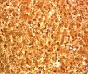
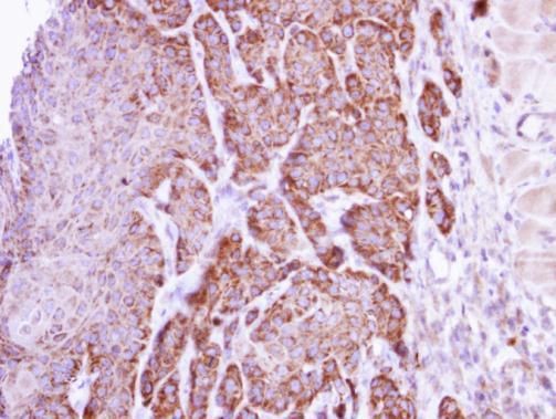
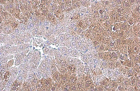
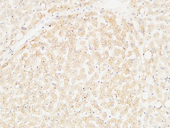
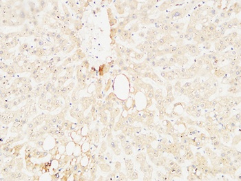
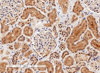
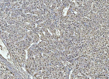
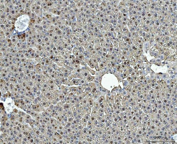
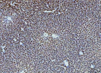
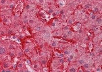
Immunohistochemical staining of Human hepatocellular carcinoma using Arginase 1 antibody
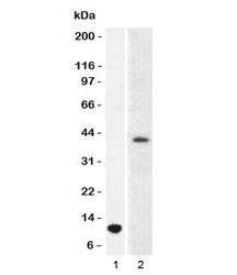
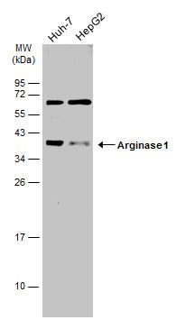
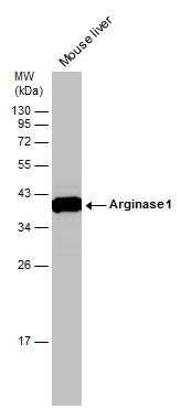
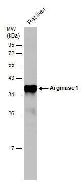
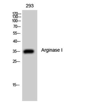
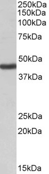
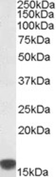
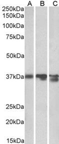
Western blot analysis of partial recombinant ARG1 protein(lane 1) and human liver(lane 2) lysate using Arginase 1 antibody
- Item 1 of 12
arginase 1 Antibody [orb556594]
ELISA, FACS, ICC, IHC-Fr, IHC-P, IP, WB
Human, Mouse, Rat
Rabbit
Polyclonal
Unconjugated
100 μl - Item 1 of 4
- Item 1 of 4
ARG1 Antibody [orb1249601]
ELISA, IHC, WB
Bovine, Canine
Human, Mouse, Porcine, Rat
Goat
Polyclonal
Unconjugated
0.1 mg - Item 1 of 4
liver Arginase/ARG1 Antibody [orb546304]
ELISA, IHC, WB
Human, Monkey, Mouse, Rat
Rabbit
Polyclonal
Unconjugated
10 μg, 100 μg - Item 1 of 3
ARG1 antibody [orb20124]
ELISA, IHC, WB
Bovine, Canine, Human, Mouse, Porcine, Rat
Goat
Polyclonal
Unconjugated
100 μg
Submit a review
Filter by Rating

- 5 stars

- 4 stars
- 3 stars
- 2 stars
- 1 stars