You have no items in your shopping cart.
Featured
Description
Research Area
Metabolism Research
Images & Validation
−

Item 1 of 5
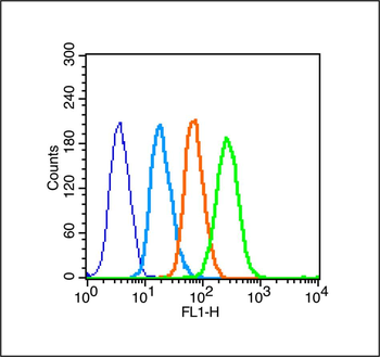
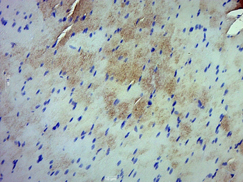
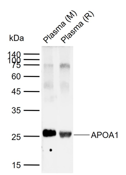
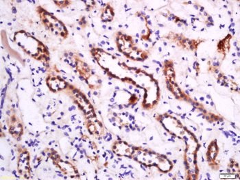
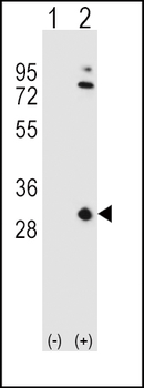
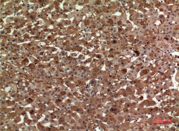
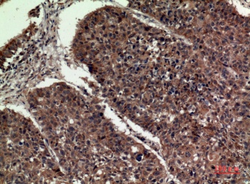
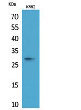
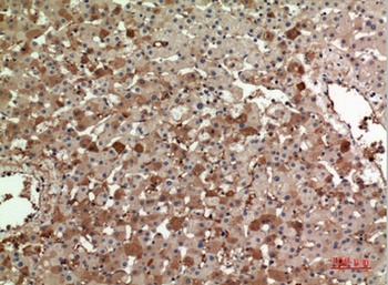
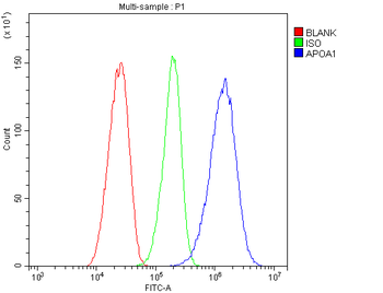
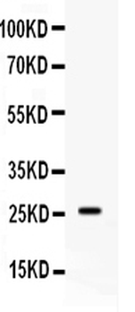
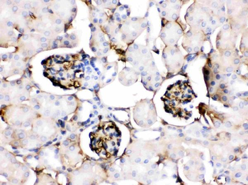
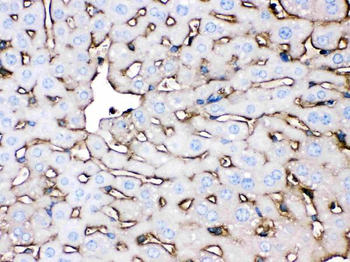
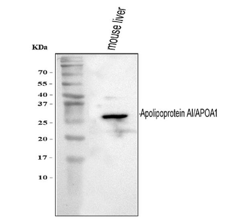
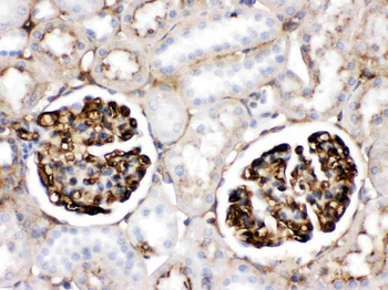
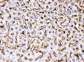
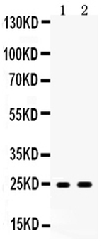
| Tested Applications | WB |
|---|---|
| Dilution Range | WB=1:500-2000 |
| Reactivity | Mouse, Rat |
| Predicted Reactivity | Human |
Key Properties
−| Antibody Type | Primary Antibody |
|---|---|
| Host | Rabbit |
| Clonality | Polyclonal |
| Isotype | IgG |
| Immunogen | KLH conjugated synthetic peptide derived from human APOA1 (51-150/267aa) |
| Target | APOA1 |
| Molecular Weight | 27 kDa |
| Purification | Affinity purified by Protein A |
| Conjugation | Unconjugated |
Storage & Handling
−| Storage | Maintain refrigerated at 2-8°C for up to 2 weeks. For long term storage store at -20°C in small aliquots to prevent freeze-thaw cycles. |
|---|---|
| Form/Appearance | Liquid |
| Buffer/Preservatives | 0.01M TBS (pH7.4) with 1% rAlbumin, 0.02% Proclin300 and 50% Glycerol. |
| Concentration | 1mg/ml |
| Expiration Date | 12 months from date of receipt. |
| Disclaimer | For research use only |
Alternative Names
−AMYLD3; HPALP2; apo(a); APOA1_HUMAN; APOA1; Apo-AI; ApoA-I; Apolipoprotein A1; apolipoprotein A-I
Similar Products
−- Item 1 of 5
- Item 1 of 4
- Item 1 of 4
Apolipoprotein A I/APOA1 Rabbit Polyclonal Antibody [orb371650]
FC, ICC, IHC, WB
Human
Rabbit
Polyclonal
Unconjugated
100 μg - Item 1 of 3
Apolipoprotein A I/APOA1 Rabbit Polyclonal Antibody [orb334577]
IHC, WB
Mouse
Rabbit
Polyclonal
Unconjugated
100 μg - Item 1 of 3
Apolipoprotein A I/APOA1 Rabbit Polyclonal Antibody [orb334578]
IHC, WB
Rat
Rabbit
Polyclonal
Unconjugated
100 μg


Quality Guarantee
Explore bioreagents carefree to elevate your research. All our products are rigorously tested for performance. If a product does not perform as described on its datasheet, our scientific support team will provide expert troubleshooting, a prompt replacement, or a refund. For full details, please see our Terms & Conditions and Buying Guide. Contact us at [email protected].
Quick Database Links
Gene Symbol
APOA1
UniProt
UniProt Details
− No UniProt data available
Bing Liang Alvin Chew1,2, An Qi Ngoh3 , Wint Wint Phoo3 , Mei Jie Grace Weng1,2, Ho Jun Sheng4 , Kitti Wing Ki Chan3 , Eddie Yong Jun Tan2,4, Terri Gelbart5 , Chenrui Xu2,4, Gene S. Tan5,6, Subhash G. Vasudevan3,7,8 & Dahai Luo Structural basis of Zika virus NS1 multimerization and human antibody recognition npj | viruses, (2024)
Available Sizes
Select a size below
Free Secondary Antibody (20 ul)0/0
Please add an antibody product to your cart first.