You have no items in your shopping cart.
Cart summary
Item 1 of 9
Item 1 of 9
Anti-TJP2/ZO2 Antibody
Catalog Number: orb865735

| Catalog Number | orb865735 |
|---|---|
| Category | Antibodies |
| Description | Anti-TJP2/ZO2 Antibody. Tested in IF, IHC, ICC, WB applications. This antibody reacts with Human, Rat, Monkey. |
| Clonality | Polyclonal |
| Species/Host | Rabbit |
| Isotype | Rabbit IgG |
| Conjugation | Unconjugated |
| Reactivity | Human, Monkey, Rat |
| Form/Appearance | Lyophilized |
| Concentration | Adding 0.2 ml of distilled water will yield a concentration of 500 μg/ml. |
| Purification | Immunogen affinity purified. |
| Immunogen | A synthetic peptide corresponding to a sequence at the C-terminus of human TJP2/ZO2. |
| UniProt ID | Q9UDY2 |
| MW | 150-170 kDa |
| Tested applications | ICC, IF, IHC, WB |
| Application notes | Western blot, 0.25-0.5 μg/ml, Human, Rat, Monkey Immunohistochemistry(Paraffin-embedded Section), 2-5 μg/ml, Human Immunocytochemistry/Immunofluorescence, 5 μg/ml, Human. Adding 0.2 ml of distilled water will yield a concentration of 500 μg/ml |
| Cross Reactivity | No cross-reactivity with other proteins. |
| Antibody Type | Primary Antibody |
| Storage | Maintain refrigerated at 2-8°C for up to 2 weeks. For long term storage store at -20°C in small aliquots to prevent freeze-thaw cycles. |
| Alternative names | CD3 antigen, epsilon polypeptide; CD3e molecule; C Read more... |
| Note | For research use only |
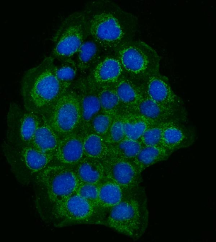
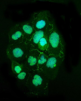
IF analysis of TJP2/ZO2 using anti-TJP2/ZO2 antibody. TJP2/ZO2 was detected in an immunocytochemical section of A431 cells. Enzyme antigen retrieval was performed using IHC enzyme antigen retrieval reagent for 15 mins. The cells were blocked with 10% goat serum. And then incubated with 5 µg/mL rabbit anti-TJP2/ZO2 Antibody overnight at 4°C. DyLight®488 Conjugated Goat Anti-Rabbit IgG was used as secondary antibody at 1:100 dilution and incubated for 30 minutes at 37°C. The section was counterstained with DAPI. Visualize using a fluorescence microscope and filter sets appropriate for the label used.
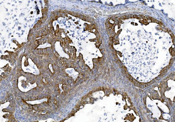
IHC analysis of TJP2/ZO2 using anti-TJP2/ZO2 antibody. TJP2/ZO2 was detected in a paraffin-embedded section of human bladder cancer tissue. Heat mediated antigen retrieval was performed in EDTA buffer (pH8.0, epitope retrieval solution). The tissue section was blocked with 10% goat serum. The tissue section was then incubated with 2 µg/ml rabbit anti-TJP2/ZO2 Antibody overnight at 4°C. Biotinylated goat anti-rabbit IgG was used as secondary antibody and incubated for 30 minutes at 37°C. The tissue section was developed using Strepavidin-Biotin-Complex (SABC) with DAB as the chromogen.
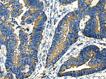
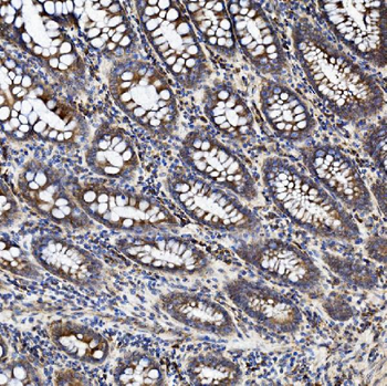
IHC analysis of TJP2/ZO2 using anti-TJP2/ZO2 antibody. TJP2/ZO2 was detected in a paraffin-embedded section of human differentiated adenocarcinoma of the rectum tissue. Heat mediated antigen retrieval was performed in EDTA buffer (pH8.0, epitope retrieval solution). The tissue section was blocked with 10% goat serum. The tissue section was then incubated with 2 µg/ml rabbit anti-TJP2/ZO2 Antibody overnight at 4°C. Biotinylated goat anti-rabbit IgG was used as secondary antibody and incubated for 30 minutes at 37°C. The tissue section was developed using Strepavidin-Biotin-Complex (SABC) with DAB as the chromogen.
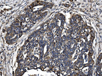
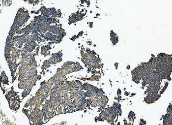
IHC analysis of TJP2/ZO2 using anti-TJP2/ZO2 antibody. TJP2/ZO2 was detected in a paraffin-embedded section of human infiltrating adenocarcinoma of the lung tissue. Heat mediated antigen retrieval was performed in EDTA buffer (pH8.0, epitope retrieval solution). The tissue section was blocked with 10% goat serum. The tissue section was then incubated with 2 µg/ml rabbit anti-TJP2/ZO2 Antibody overnight at 4°C. Biotinylated goat anti-rabbit IgG was used as secondary antibody and incubated for 30 minutes at 37°C. The tissue section was developed using Strepavidin-Biotin-Complex (SABC) with DAB as the chromogen.
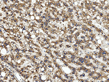
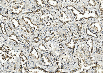
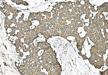
IHC analysis of TJP2/ZO2 using anti-TJP2/ZO2 antibody. TJP2/ZO2 was detected in a paraffin-embedded section of human liver cancer tissue. Heat mediated antigen retrieval was performed in EDTA buffer (pH8.0, epitope retrieval solution). The tissue section was blocked with 10% goat serum. The tissue section was then incubated with 2 µg/ml rabbit anti-TJP2/ZO2 Antibody overnight at 4°C. Biotinylated goat anti-rabbit IgG was used as secondary antibody and incubated for 30 minutes at 37°C. The tissue section was developed using Strepavidin-Biotin-Complex (SABC) with DAB as the chromogen.
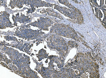
IHC analysis of TJP2/ZO2 using anti-TJP2/ZO2 antibody. TJP2/ZO2 was detected in a paraffin-embedded section of human ovarian serous adenocarcinoma tissue. Heat mediated antigen retrieval was performed in EDTA buffer (pH8.0, epitope retrieval solution). The tissue section was blocked with 10% goat serum. The tissue section was then incubated with 2 µg/ml rabbit anti-TJP2/ZO2 Antibody overnight at 4°C. Biotinylated goat anti-rabbit IgG was used as secondary antibody and incubated for 30 minutes at 37°C. The tissue section was developed using Strepavidin-Biotin-Complex (SABC) with DAB as the chromogen.
IHC analysis of TJP2/ZO2 using anti-TJP2/ZO2 antibody. TJP2/ZO2 was detected in a paraffin-embedded section of human papillary carcinoma of the left breast tissue. Heat mediated antigen retrieval was performed in EDTA buffer (pH8.0, epitope retrieval solution). The tissue section was blocked with 10% goat serum. The tissue section was then incubated with 2 µg/ml rabbit anti-TJP2/ZO2 Antibody overnight at 4°C. Biotinylated goat anti-rabbit IgG was used as secondary antibody and incubated for 30 minutes at 37°C. The tissue section was developed using Strepavidin-Biotin-Complex (SABC) with DAB as the chromogen.
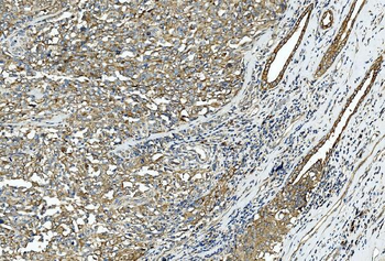
IHC analysis of TJP2/ZO2 using anti-TJP2/ZO2 antibody. TJP2/ZO2 was detected in a paraffin-embedded section of human squamous metaplasia of the renal pelvis tissue. Heat mediated antigen retrieval was performed in EDTA buffer (pH8.0, epitope retrieval solution). The tissue section was blocked with 10% goat serum. The tissue section was then incubated with 2 µg/ml rabbit anti-TJP2/ZO2 Antibody overnight at 4°C. Biotinylated goat anti-rabbit IgG was used as secondary antibody and incubated for 30 minutes at 37°C. The tissue section was developed using Strepavidin-Biotin-Complex (SABC) with DAB as the chromogen.
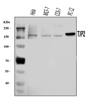
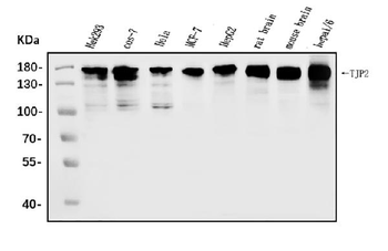
Western blot analysis of TJP2/ZO2 using anti-TJP2/ZO2 antibody. Electrophoresis was performed on a 5-20% SDS-PAGE gel at 70V (Stacking gel) / 90V (Resolving gel) for 2-3 hours. The sample well of each lane was loaded with 30 ug of sample under reducing conditions. Lane 1: human Hela whole cell lysates, Lane 2: human MCF-7 whole cell lysates, Lane 3: monkey COS-7 whole cell lysates, Lane 4: rat PC-12 whole cell lysates. After electrophoresis, proteins were transferred to a nitrocellulose membrane at 150 mA for 50-90 minutes. Blocked the membrane with 5% non-fat milk/TBS for 1.5 hour at RT. The membrane was incubated with rabbit anti-TJP2/ZO2 antigen affinity purified polyclonal antibody at 0.5 µg/mL overnight at 4°C, then washed with TBS-0.1% Tween 3 times with 5 minutes each and probed with a goat anti-rabbit IgG-HRP secondary antibody at a dilution of 1:5000 for 1.5 hour at RT. The signal is developed using an Enhanced Chemiluminescent detection (ECL) kit with Tanon 5200 system. A specific band was detected for TJP2/ZO2 at approximately 150-170 kDa. The expected band size for TJP2/ZO2 is at 134 kDa.
- Item 1 of 6
Anti-TJP2/ZO2 Antibody [orb745930]
FC, ICC, IF, IHC, WB
Human, Monkey, Mouse, Rat
Rabbit
Polyclonal
Unconjugated
100 μg, 10 μg 
Anti-TJP2/ZO2 Antibody [orb2602109]
FC, ICC, IF, IHC, WB
Human, Monkey, Mouse, Rat
Rabbit
Polyclonal
iFluor647
100 μgAnti-TJP2/ZO2 Antibody [orb2602110]
FC, ICC, IF, IHC, WB
Human, Monkey, Mouse, Rat
Rabbit
Polyclonal
PE
100 μgAnti-TJP2/ZO2 Antibody [orb2602111]
FC, ICC, IF, IHC, WB
Human, Monkey, Mouse, Rat
Rabbit
Polyclonal
APC
100 μgAnti-TJP2/ZO2 Antibody [orb2602112]
FC, ICC, IF, IHC, WB
Human, Monkey, Mouse, Rat
Rabbit
Polyclonal
HRP
100 μg