You have no items in your shopping cart.
Featured
Description
Research Area
Cell Biology
Images & Validation
−Item 1 of 6
| Tested Applications | FC, ICC, IHC-P, WB |
|---|---|
| Reactivity | Canine, Human, Mouse, Porcine, Rat |
| Application Notes |
Key Properties
−| Antibody Type | Primary Antibody |
|---|---|
| Clonality | Monoclonal |
| Isotype | Mouse IgG1 |
| Clone No. | TU-20 |
| Immunogen | Peptide (C) 441-448 coupled to maleimide-activated keyhole limpet hemocyanin via cysteine added to the N-terminus of the neuron-specific peptide. |
| Target | betaIII-Tubulin |
| Purification | Purified by protein-A affinity chromatography. |
| Conjugation | Unconjugated |
Storage & Handling
−| Storage | Maintain refrigerated at 2-8°C for up to 2 weeks. For long term storage store at -20°C in small aliquots to prevent freeze-thaw cycles. |
|---|---|
| Buffer/Preservatives | Phosphate buffered saline (PBS), pH 7.4, 15 mM sodium azide |
| Concentration | 1 mg/ml |
| Expiration Date | 12 months from date of receipt. |
| Disclaimer | For research use only |
Alternative Names
−TUBB3
Similar Products
−- Item 1 of 2
betaIII-Tubulin Antibody (FITC) [orb44545]
FC, ICC
Canine, Human, Mouse, Porcine, Rat
Monoclonal
FITC
0.1 mg - Item 1 of 2
Chicken Tubulin beta 3 Antibody [orb3011859]
IF, IHC-Fr, WB
Human, Mouse, Rat
Gallus
Unconjugated
50 μl, 100 μl - Item 1 of 2
Guinea pig anti-Tubulin beta 3 Antibody [orb2806691]
IF, IHC-Fr, WB
Human, Mouse, Rat
Guinea pig
Unconjugated
50 μl, 100 μl - Item 1 of 2
Goat anti-Tubulin beta 3 Antibody [orb2806731]
IF, IHC-Fr, WB
Human, Mouse, Rat
Goat
Unconjugated
50 μl, 100 μl


Quality Guarantee
Explore bioreagents carefree to elevate your research. All our products are rigorously tested for performance. If a product does not perform as described on its datasheet, our scientific support team will provide expert troubleshooting, a prompt replacement, or a refund. For full details, please see our Terms & Conditions and Buying Guide. Contact us at [email protected].
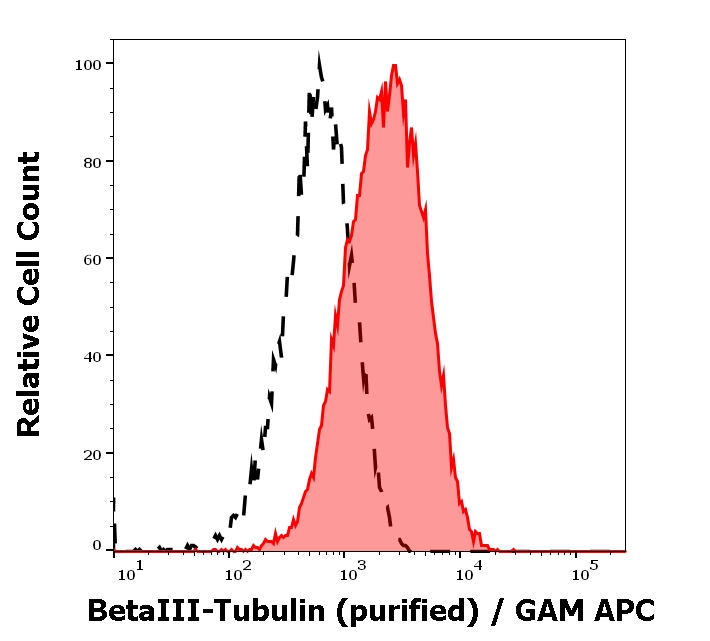
Separation of MCF-7 cells stained using anti-betaIII-Tubulin (TU-20) purified antibody (concentration in sample 9 µg/ml, GAM APC, red-filled) from MCF-7 cells unstained by primary antibody (GAM APC, black-dashed) in flow cytometry analysis (intracellular staining).
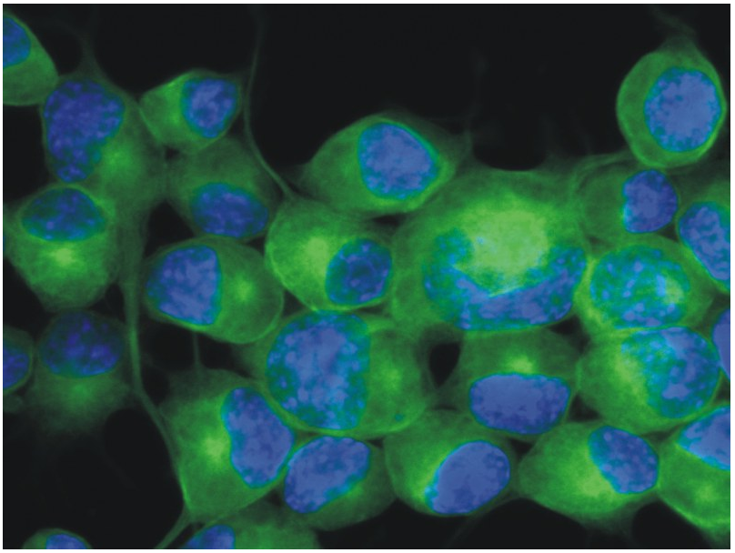
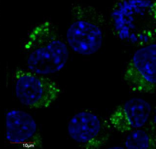
Immunocytochemistry staining of Neuro2a mouse neuroblastoma cell line using anti-betaIII-tubulin (TU-20; green; 3 µg/ml). Nuclei were stained with DAPI (blue).
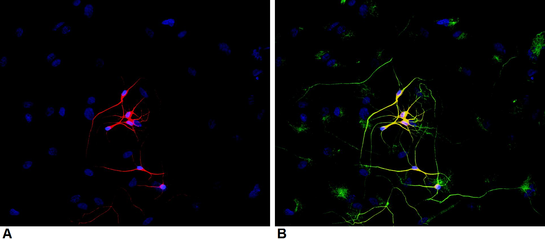
Immunocytochemistry staining of P-19 mouse embryonal carcinoma cell line stimulated to neuronal differentiation by retinoic acid. A - Microtubules decorated with neuron-specific anti-betaIII-tubulin (TU-20; red). B - Merged image of co-staining with anti-beta-tubulin (TU-06; green; cat. no. orb44535). Superposition of red and green colours provided yellow staining. Nuclei were stained with DNA-binding dye (blue).
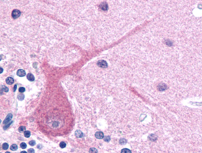
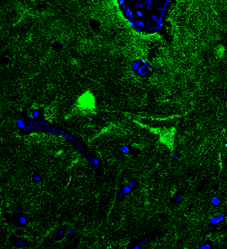
Immunohistochemistry staining of human brain (paraffin sections) using anti-betaIII tubulin (TU-20).
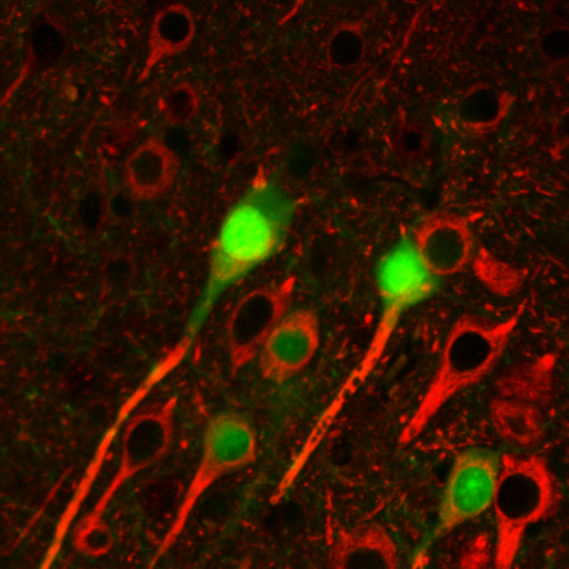
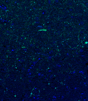
Immunohistochemistry staining of betaIII tubulin (red) in tissue sections of murine brain expressing GFP in some of its neurons (green). Mouse monoclonal antibody TU-20 (anti-betaIII tubulin) was detected with goat anti-mouse IgG1 conjugated with Alexa Fluor 555.
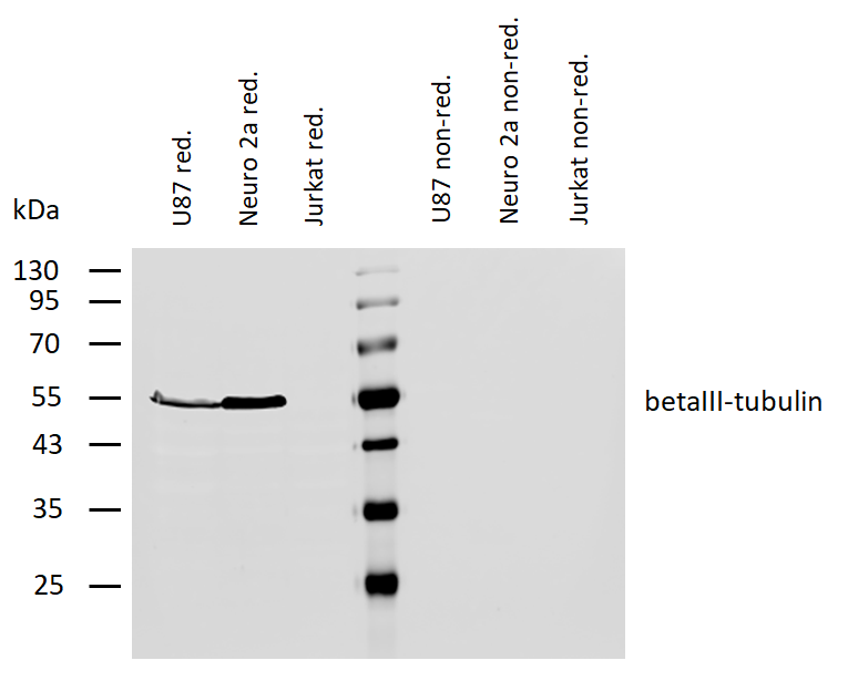
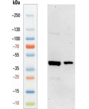
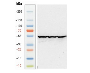
Western blotting analysis of human betaIII-tubulin using mouse monoclonal antibody TU-20 on lysates of U87 cells and Neuro 2a cells (and Jurkat cells as a negative control) under reducing and non-reducing conditions. Nitrocellulose membrane was probed with 2 µg/ml of mouse anti-betaIII-tubulin monoclonal antibody followed by IRDye800-conjugated anti-mouse secondary antibody. A specific band was detected for betaIII-tubulin at approximately 55 kDa.
Documents Download
Datasheet
Product Information
Request a Document
Protocol Information
WB
Western Blot (IB, immunoblot)
IHC-P
Immunohistochemistry Paraffin
FC
Flow Cytometry
ICC
Immunocytochemistry
Sana, Jiri et al. Identification of microRNAs differentially expressed in glioblastoma stem-like cells and their association with patient survival Sci Rep, 8, 2836 (2018)
betaIII-Tubulin Antibody (orb44544)

- 0.0
Based on 0 reviews
Participating in our Biorbyt product reviews program enables you to support fellow scientists by sharing your firsthand experience with our products.
Login to Submit a ReviewAvailable Sizes
Select a size below
Free Secondary Antibody (20 ul)0/0
Please add an antibody product to your cart first.