You have no items in your shopping cart.
Cart summary
Item 1 of 5
Item 1 of 5
ALDH1A1 Antibody
Catalog Number: orb1268148

| Catalog Number | orb1268148 |
|---|---|
| Category | Antibodies |
| Description | ALDH1A1 Antibody |
| Target | ALDH1A1 |
| Clonality | Polyclonal |
| Isotype | Rabbit Ig |
| Conjugation | Unconjugated |
| Reactivity | Human |
| Predicted Reactivity | Monkey, Mouse |
| Form/Appearance | Liquid |
| Concentration | batch dependent |
| Buffer/Preservatives | Supplied in PBS with 0.09% (W/V) sodium azide. |
| Purification | This antibody is prepared by Saturated Ammonium Sulfate (SAS) precipitation followed by dialysis |
| Immunogen | This ALDH1A1 antibody is generated from rabbits immunized with a KLH conjugated synthetic peptide between 302-331 amino acids from the Central region of human ALDH1A1. |
| UniProt ID | P00352 |
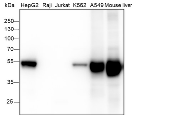
| MW | 55 kDa |
| Tested applications | FC, IF, IHC-P, WB |
| Application notes | For WB starting dilution is: 1:1000For IHC-P starting dilution is: 1:10~50For IF starting dilution is: 1:10~50For FACS starting dilution is: 1:10~50 |
| Antibody Type | Primary Antibody |
| Storage | Maintain refrigerated at 2-8°C for up to 2 weeks. For long term storage store at -20°C in small aliquots to prevent freeze-thaw cycles. |
| Alternative names | Retinal dehydrogenase 1, RALDH 1, RalDH1, ALDH-E1, Read more... |
| Note | For research use only |
| NCBI | P00352 |
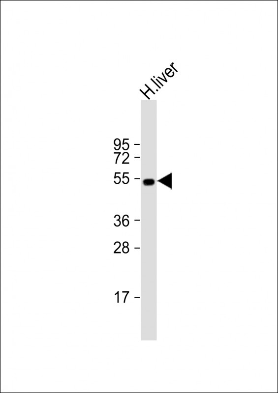
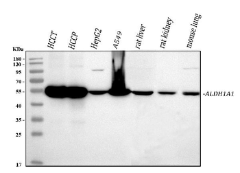
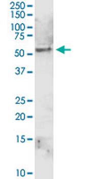
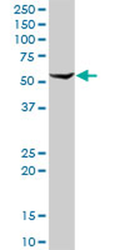
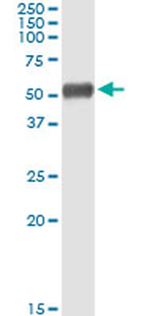
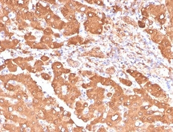
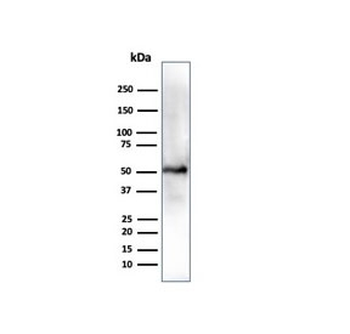
Western Blot at 1:1000 dilution + human liver lysate Lysates/proteins at 20 ug per lane.
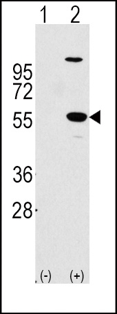
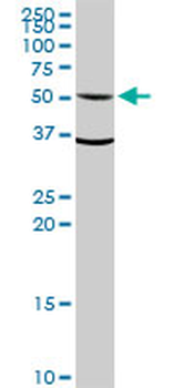
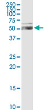
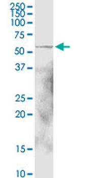
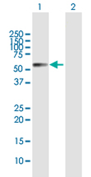
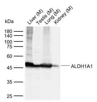
Western blot analysis of ALDH1A1 using rabbit polyclonal ALDH1A1 Antibody using 293 cell lysates (2 ug/lane) either nontransfected (Lane 1) or transiently transfected with the ALDH1A1 gene (Lane 2).
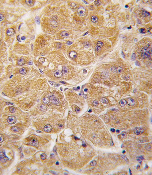
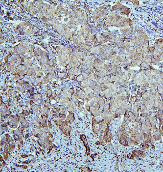
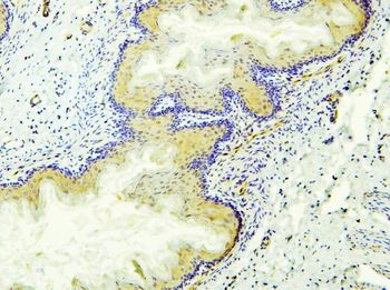
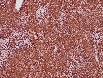
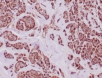
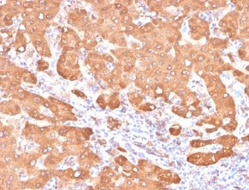
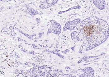
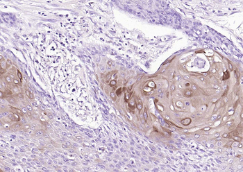
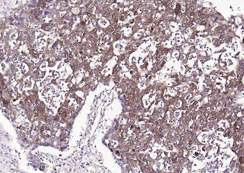
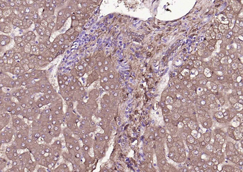
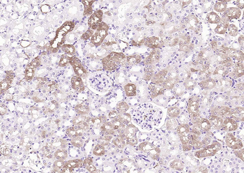
Formalin-fixed and paraffin-embedded human hepatocarcinoma tissue reacted with ALDH1A1 antibody, which was peroxidase-conjugated to the secondary antibody, followed by DAB staining.
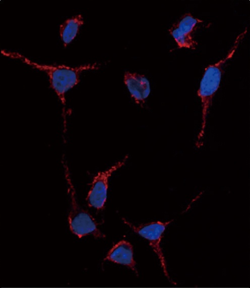
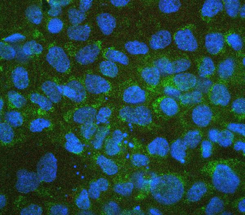
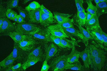
Immunofluorescence analysis of anti-ALDH1A1 Antibody in HeLa cells. 0.025 mg/ml primary antibody was followed by Alexa-Fluor-546-conjugated donkey anti-rabbit lgG (H + L). Alexa-Fluor-546 emits orange fluorescence. Blue counterstaining is DAPI.
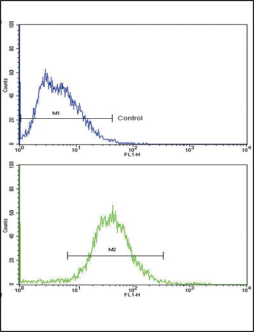
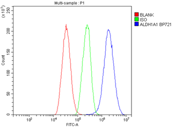
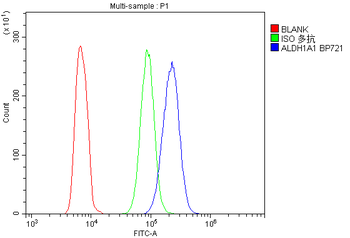
Flow cytometric analysis of HepG2 cells using ALDH1A1 Antibody (bottom histogram) compared to a negative control cell (top histogram). FITC-conjugated goat-anti-rabbit secondary antibodies were used for the analysis.
- Item 1 of 10
Retinal dehydrogenase 1 ALDH1A1 Antibody [orb570330]
ELISA, FC, ICC, IF, IHC, WB
Human, Mouse, Rat
Rabbit
Polyclonal
Unconjugated
100 μg, 10 μg - Item 1 of 7
ALDH1A1 MaxPab rabbit polyclonal antibody (D01) [orb2296206]
IP, WB
Human, Mouse
Rabbit
Polyclonal
Unconjugated
100 μl - Item 1 of 7
Aldehyde Dehydrogenase 1A1 Antibody / ALDH1A1 [orb2635093]
IHC-P, WB
Human
Mouse
Monoclonal
Unconjugated
100 μg - Item 1 of 7
Aldehyde Dehydrogenase 1A1 Antibody / ALDH1A1 [orb2635094]
IHC-P, WB
Human
Mouse
Monoclonal
Unconjugated
20 μg, 100 μg - Item 1 of 7
ALDH1A1 Recombinant Rabbit Monoclonal Antibody [orb1152015]
IF, IHC-Fr, IHC-P, WB
Mouse
Human, Mouse
Rabbit
Recombinant
Unconjugated
25 μl, 100 μl, 50 μl