You have no items in your shopping cart.
Cart summary
Item 1 of 6
Item 1 of 6
AHSG Antibody
Catalog Number: orb1270962

| Catalog Number | orb1270962 |
|---|---|
| Category | Antibodies |
| Description | AHSG Antibody |
| Target | AHSG |
| Clonality | Polyclonal |
| Isotype | Rabbit Ig |
| Conjugation | Unconjugated |
| Reactivity | Human |
| Form/Appearance | Liquid |
| Concentration | batch dependent |
| Buffer/Preservatives | Supplied in PBS with 0.09% (W/V) sodium azide. |
| Purification | This antibody is purified through a protein A column, followed by peptide affinity purification. |
| Immunogen | This AHSG antibody is generated from rabbits immunized with a KLH conjugated synthetic peptide between 247-276 amino acids from the C-terminal region of human AHSG. |
| UniProt ID | P02765 |
| MW | 39 kDa |
| Tested applications | FC, IF, IHC-P, WB |
| Application notes | For FACS starting dilution is: 1:25For IF starting dilution is: 1:25For IHC-P starting dilution is: 1:25For WB starting dilution is: 1:1000 |
| Antibody Type | Primary Antibody |
| Storage | Maintain refrigerated at 2-8°C for up to 2 weeks. For long term storage store at -20°C in small aliquots to prevent freeze-thaw cycles. |
| Alternative names | Alpha-2-HS-glycoprotein, Alpha-2-Z-globulin, Ba-al Read more... |
| Note | For research use only |
| NCBI | P02765 |

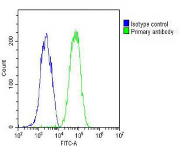
Overlay histogram showing HepG2 cells stained with Antibody (green line). The cells were fixed with 2% paraformaldehyde (10 min) and then permeabilized with 90% methanol for 10 min. The cells were then icubated in 2% bovine serum albumin to block non-specific protein-protein interactions followed by the antibody (1:25 dilution) for 60 min at 37oC. The secondary antibody used was Goat-Anti-Rabbit IgG, DyLight 488 Conjugated Highly Cross-Adsorbed (OH191631) at 1/200 dilution for 40 min at 37oC. Isotype control antibody (blue line) was rabbit IgG (1 ug/1x10^6 cells) used under the same conditions. Acquisition of >10000 events was performed.
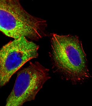
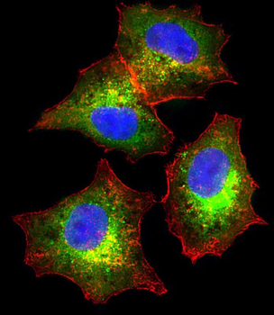
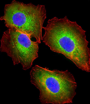
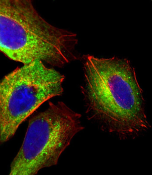
Immunofluorescent analysis of 4% paraformaldehyde-fixed, 0. 1% Triton X-100 permeabilized HepG2 (human liver hepatocellular carcinoma cell line) cells labeling Pdx1 with antibody at 1/25 dilution, followed by Dylight 488-conjugated goat anti-rabbit IgG (NK179883) secondary antibody at 1/200 dilution (green). Immunofluorescence image showing cytoplasm staining on HepG2 cell line. Cytoplasmic actin is detected with Dylight 554 Phalloidin (PD18466410) at 1/100 dilution (red). The nuclear counter stain is DAPI (blue).
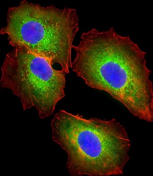
Immunofluorescent analysis of 4% paraformaldehyde-fixed, 0. 1% Triton X-100 permeabilized HepG2 (human liver hepatocellular carcinoma cell line) cells labeling Pdx1 with antibody at 1/25 dilution, followed by Dylight 488-conjugated goat anti-rabbit IgG (NK179883) secondary antibody at 1/200 dilution (green). Immunofluorescence image showing cytoplasm staining on HepG2 cell line. Cytoplasmic actin is detected with Dylight 554 Phalloidin (PD18466410) at 1/100 dilution (red). The nuclear counter stain is DAPI (blue).

Immunofluorescent analysis of 4% paraformaldehyde-fixed, 0. 1% Triton X-100 permeabilized HepG2 (human liver hepatocellular carcinoma cell line) cells labeling Pdx1 with antibody at 1/25 dilution, followed by Dylight 488-conjugated goat anti-rabbit IgG (NK179883) secondary antibody at 1/200 dilution (green). Immunofluorescence image showing cytoplasm staining on HepG2 cell line. Cytoplasmic actin is detected with Dylight 554 Phalloidin (PD18466410) at 1/100 dilution (red). The nuclear counter stain is DAPI (blue).

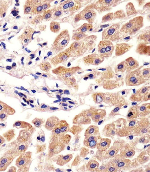
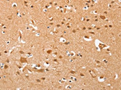
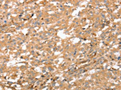
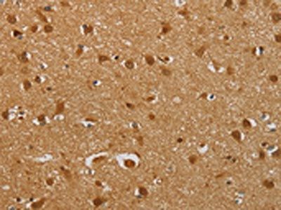
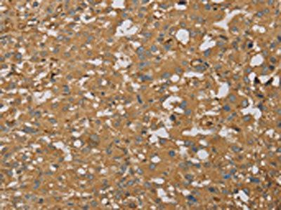
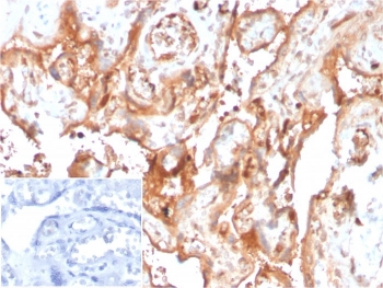
Antibody staining AHSG in human liver tissue sections by Immunohistochemistry (IHC-P - paraformaldehyde-fixed, paraffin-embedded sections).
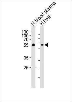
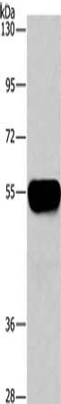
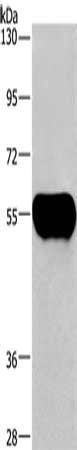
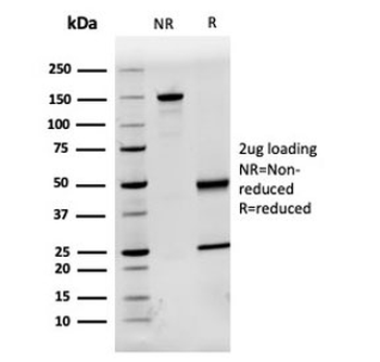
Western blot analysis of lysates from human blood plasma and liver tissue lysates (from left to right), using AHSG Antibody at 1:1000 at each lane.
- Item 1 of 6
AHSG Antibody (C-term) [orb1939166]
FC, IF, IHC-P, WB
Human
Rabbit
Polyclonal
Unconjugated
100 μl, 50 μl - Item 1 of 3
- Item 1 of 3
- Item 1 of 3
- Item 1 of 3