You have no items in your shopping cart.
Cart summary
Item 1 of 4
Item 1 of 4
AGL Antibody
Catalog Number: orb1264727

| Catalog Number | orb1264727 |
|---|---|
| Category | Antibodies |
| Description | AGL Antibody |
| Clonality | Polyclonal |
| Tested applications | IF, WB |
| Reactivity | Human |
| Isotype | Rabbit Ig |
| Immunogen | This AGL antibody is generated from rabbits immunized with a KLH conjugated synthetic peptide between 1479-1510 amino acids from the C-terminal region of human AGL. |
| Antibody Type | Primary Antibody |
| Concentration | batch dependent |
| Form/Appearance | Liquid |
| Conjugation | Unconjugated |
| MW | 175 kDa |
| Target | AGL |
| UniProt ID | P35573 |
| NCBI | P35573 |
| Storage | Maintain refrigerated at 2-8°C for up to 2 weeks. For long term storage store at -20°C in small aliquots to prevent freeze-thaw cycles. |
| Buffer/Preservatives | Supplied in PBS with 0.09% (W/V) sodium azide. |
| Alternative names | Glycogen debranching enzyme, Glycogen debrancher, Read more... |
| Note | For research use only |
| Application notes | For WB starting dilution is: 1:1000For IF starting dilution is: 1:10~50 |
| Expiration Date | 12 months from date of receipt. |
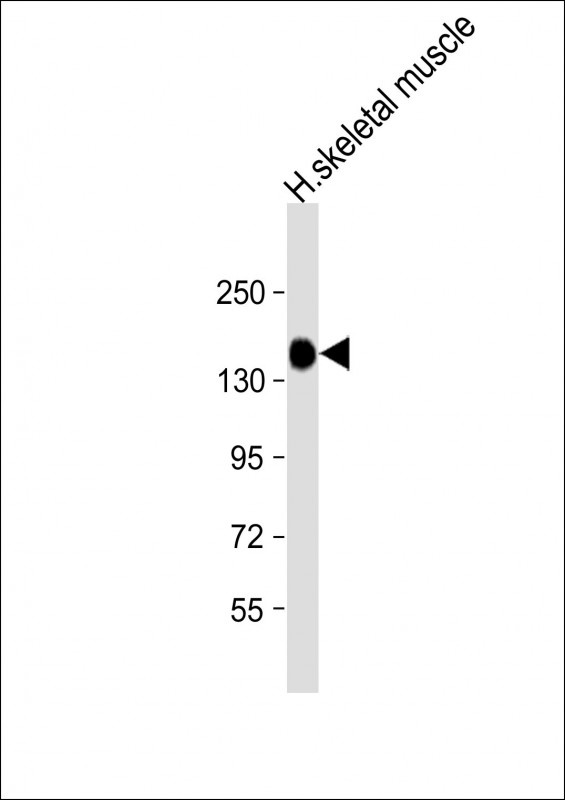
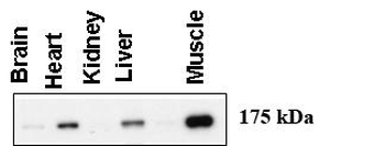
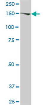
Western Blot at 1:8000 dilution + human skeletal muscle lysate Lysates/proteins at 20 ug per lane.

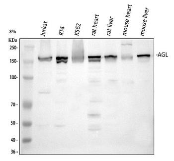
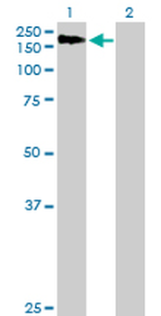
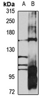
Western blot using anti-AGL antibody at 1:1000 dilution. A total of 20 ug of lysates was loaded for each tissue.

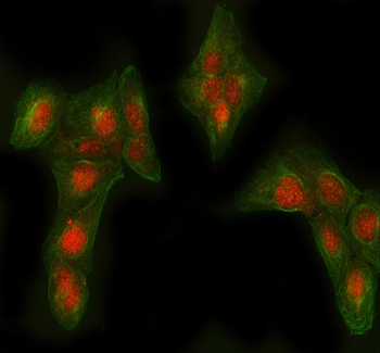
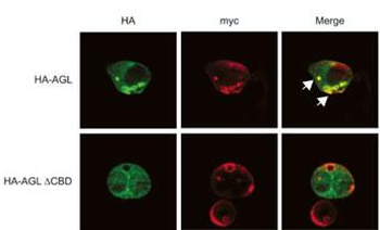
Expression of myc-GS causes wild type but not the ÃCBD mutant of AGL to aggregate around the PAS-stain-positive inclusions. HepG2 cells were transfected with either HA-tagged wild-type AGL (HA-AGL) or HA-AGL ÃCBD. Cells were fixed in formalin and processed for IF using anti-HA (green) and anti-myc (red) antibodies. White arrows indicate colocalization of HA-AGL and myc-GS.
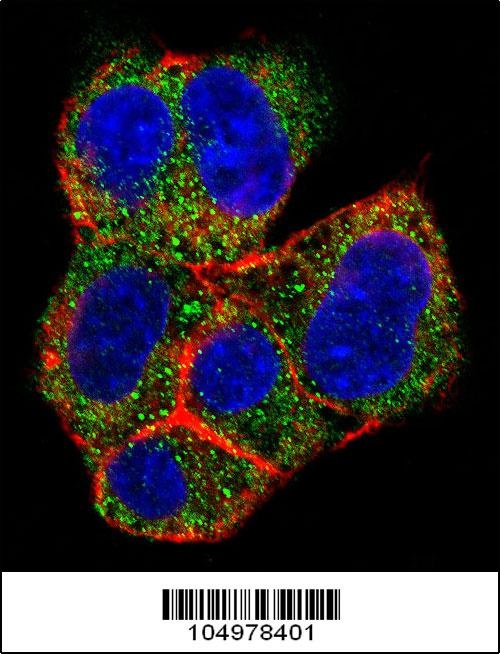
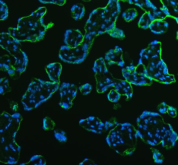
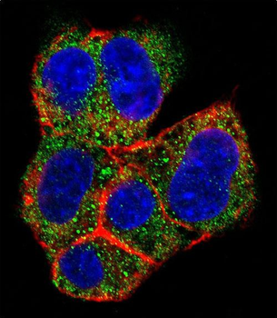
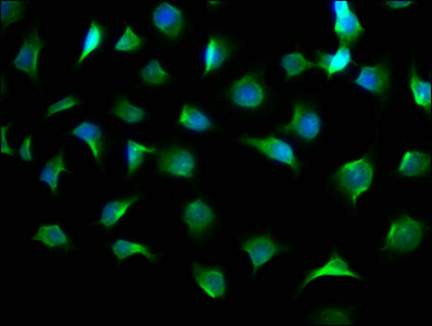
Confocal immunofluorescent analysis of AGL Antibody with HepG2 cell followed by Alexa Fluor 488-conjugated goat anti-rabbit lgG (green). Actin filaments have been labeled with Alexa Fluor 555 phalloidin (red). DAPI was used to stain the cell nuclear (blue).
- Item 1 of 5
Anti-AGL Antibody [orb1804663]
ELISA, FC, ICC, IF, IHC, WB
Human, Mouse, Rat
Rabbit
Polyclonal
Unconjugated
10 μg, 100 μg - Item 1 of 4
- Item 1 of 2
AGL purified MaxPab mouse polyclonal antibody (B01P) [orb2296299]
WB
Human
Mouse
Polyclonal
Unconjugated
50 μg - Item 1 of 1
- Item 1 of 1