You have no items in your shopping cart.
Cart summary
Item 1 of 3
Item 1 of 3
ACTA1 antibody
Catalog Number: orb213515
| Catalog Number | orb213515 |
|---|---|
| Category | Antibodies |
| Description | Rabbit polyclonal antibody against ACTA1 |
| Species/Host | Rabbit |
| Clonality | Polyclonal |
| Tested applications | IH, IP, WB |
| Reactivity | Human, Mouse, Porcine, Rabbit, Rat |
| Immunogen | KLH-conjugated synthetic peptide encompassing a sequence within the N-term region of human Alpha-actin-1. The exact sequence is proprietary. |
| Dilution range | WB: 1-500-1-1000, IHC-P: 1-100-1-200, IP: 1-10-1-100 |
| Form/Appearance | Liquid in 0.42% Potassium phosphate, 0.87% Sodium chloride, pH 7.3, 30% glycerol, and 0.01% sodium azide. |
| Conjugation | Unconjugated |
| Target | ACTA1 |
| Entrez | 11459, 58, 29437 |
| UniProt ID | P68133, P68136, P68134 |
| Source | Rabbit |
| Storage | Shipped at 4°C. Upon delivery aliquot and store at -20°C for one year. Avoid freeze/thaw cycles. |
| Buffer/Preservatives | Liquid in 0.42% Potassium phosphate, 0.87% Sodium chloride, pH 7.3, 30% glycerol, and 0.01% sodium azide. |
| Alternative names | anti-ACTA antibody, anti-ACTA1 antibody, anti-ACTA Read more... |
| Note | For research use only |
| Expiration Date | 12 months from date of receipt. |
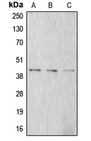
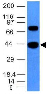
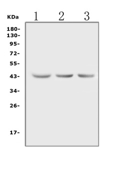
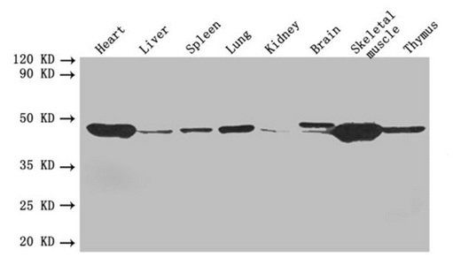
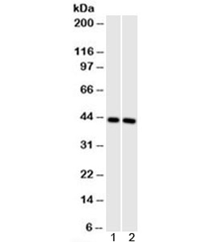
Western blot analysis of Alpha-actin-1 expression in A549 (A), SP2/0 (B), rat kidney (C) whole cell lysates. (Predicted band size: 42 kD; Observed band size: 42 kD)
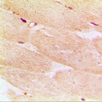
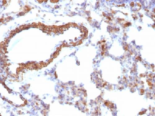
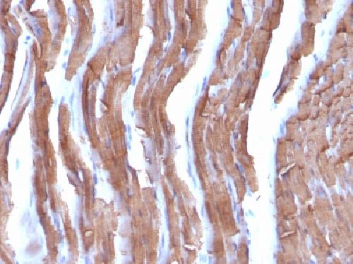
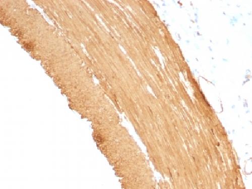
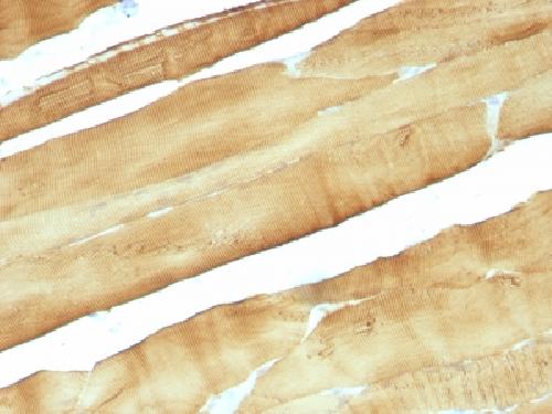
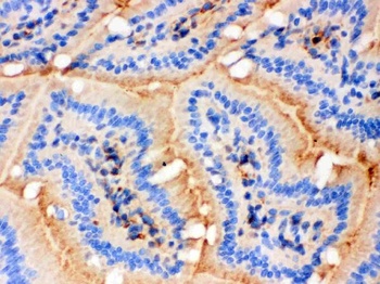
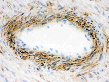
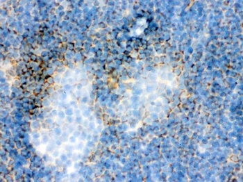
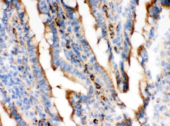
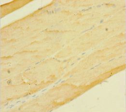
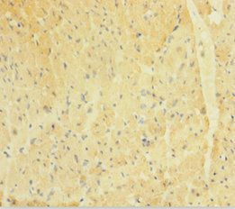
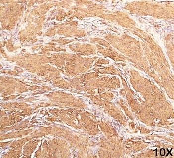
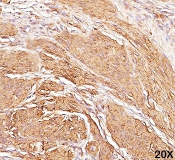
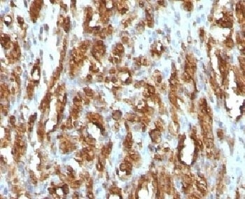
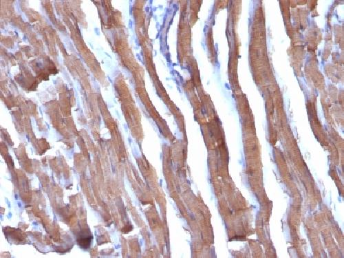
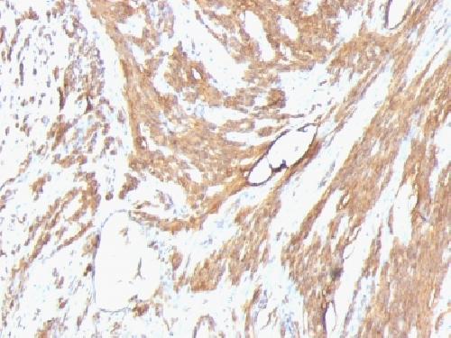
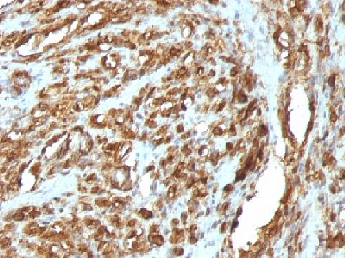
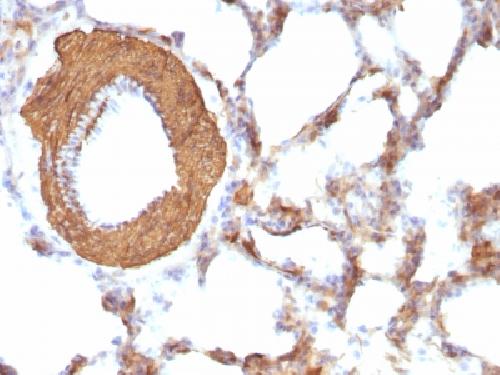
Immunohistochemical analysis of Alpha-actin-1 staining in human muscle formalin fixed paraffin embedded tissue section. The section was pre-treated using heat mediated antigen retrieval with sodium citrate buffer (pH 6.0). The section was then incubated with the antibody at room temperature and detected using an HRP conjugated compact polymer system. DAB was used as the chromogen. The section was then counterstained with haematoxylin and mounted with DPX.

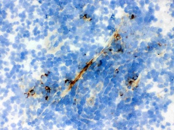
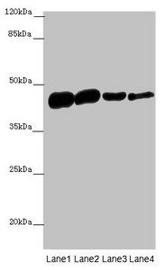
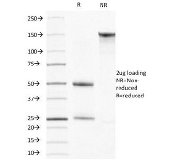
Immunoprecipitation of Alpha-actin-1 from 0.5 mg Mouse Skeletal Muscle tissue lysate, using 5 ug of Anti-Alpha-actin-1 Antibody and 50 ul of protein G magnetic beads (+). No antibody was added to the control (-). The antibody was incubated under agitation with Protein G beads for 10 min, Mouse Skeletal Muscle tissue lysate diluted in RIPA buffer was added to each sample and incubated for a further 10 min under agitation. Proteins were eluted by addition of 40 ul SDS loading buffer and incubated for 10 min at 70°C; 10 ul of each sample was separated on a SDS PAGE gel, transferred to a nitrocellulose membrane, blocked with 5% BSA and probed with Anti-Alpha-actin-1 Antibody.
- Item 1 of 7
- Item 1 of 6
Actin ACTA1 Antibody (Monoclonal, AC-40) [orb18178]
IHC, WB
Gallus, Human, Mouse, Rat
Mouse
Monoclonal
Unconjugated
10 μg, 100 μg - Item 1 of 4
ACTA1 antibody [orb241463]
ELISA, IHC, WB
Human, Mouse, Rat
Rabbit
Polyclonal
Unconjugated
50 μg, 100 μg - Item 1 of 5
ACTA1 Antibody [orb1252673]
FC, IF, IHC, WB
Human, Mouse, Rabbit, Rat
Mouse
Monoclonal
Unconjugated
100 μg - Item 1 of 5
ACTA1 antibody [orb389301]
FC, IF, IHC-P
Canine, Feline, Human, Rabbit, Rat
Mouse
Monoclonal
Unconjugated
100 μg, 20 μg

Submit a review
Filter by Rating

- 5 stars

- 4 stars
- 3 stars
- 2 stars
- 1 stars