You have no items in your shopping cart.
10X RIPA Lysis Buffer
SKU: orb420112
Description
Images & Validation
−Item 1 of 2
| Application Notes |
|---|
Key Properties
−| Purity | This product was aseptically filtered through a Millipore 0.22 micron filter into clean, pre-sterilized containers. The product was tested on trypticase soy agar for 24 hours, 48 hours and 72 hours and was found to be negative for bacteria. |
|---|---|
| Conjugation | Unconjugated |
Storage & Handling
−| Storage | Store container at room temperature (18° to 26° C) prior to opening. Protect from light (store in the dark). |
|---|---|
| Form/Appearance | Liquid (sterile filtered) |
| Buffer/Preservatives | Preservative: 0.01% (w/v) Sodium Azide. Stabilizer: None; Buffer: See application note. |
| Concentration | 10X |
| Expiration Date | 12 months from date of receipt. |
| Hazard Information | Non-Toxic |
| Disclaimer | For research use only |
Alternative Names
−10X RIPA Lysis Buffer, RIPA (Radio-Immunoprecipitation Assay) Lysis Buffer
Similar Products
−Quality Guarantee
Explore bioreagents carefree to elevate your research. All our products are rigorously tested for performance. If a product does not perform as described on its datasheet, our scientific support team will provide expert troubleshooting, a prompt replacement, or a refund. For full details, please see our Terms & Conditions and Buying Guide. Contact us at [email protected].
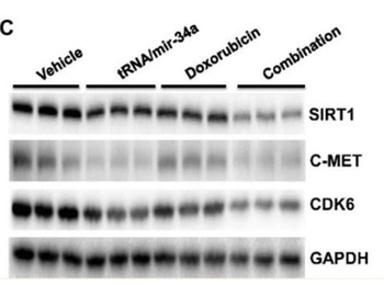
Comparison of miR-34a and target oncogene expression levels in 143B cells treated with bioengineered miR-34a prodrug (tRNA/mir-34a) and doxorubicin, alone or in combination. Cells were harvested at 72 h after treatment. Pre-miR-34a. (C) were measured by Western blot. Band density was determined by Image Lab software (Bio-Rad), and normalized to that of GAPDH. Cell lysates were prepared using RIPA buffer supplemented with the complete protease inhibitor cocktail, and protein concentrations were determined using a BCA Protein Assay Kit.
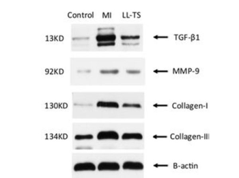
Representative picture above of Western blots from LV-free wall tissues in each group (control group, 10; MI group, 5; LL-TS group, 5) showed effects of LL-TS treatment on protein expression level of TGF-β1, MMP-9, collagen I, and collagen III. Transmural myocardial tissue sample ≈ 1 cm2 obtained from the LV free wall outside the infarction area was homogenized in radioimmunoprecipitation assay lysis buffer containing proteinase inhibitor.
Protocol Information
WB
Western Blot (IB, immunoblot)
IP
Immunoprecipitation
ChIP
Chromatin Immunoprecipitation
10X RIPA Lysis Buffer (orb420112)

- 0.0
Based on 0 reviews
Participating in our Biorbyt product reviews program enables you to support fellow scientists by sharing your firsthand experience with our products.
Login to Submit a ReviewAvailable Sizes
Select a size below