You have no items in your shopping cart.
Cart summary
Item 1 of 11
Item 1 of 11
XBP1 Antibody
Catalog Number: orb1240297
| Catalog Number | orb1240297 |
|---|---|
| Category | Antibodies |
| Description | XBP1 Antibody |
| Species/Host | Rabbit |
| Clonality | Polyclonal |
| Tested applications | ELISA, ICC, IF, IHC-P, WB |
| Predicted Reactivity | Bovine |
| Reactivity | Human, Mouse, Rat |
| Isotype | IgG |
| Immunogen | Anti-XBP-1 antibody (orb1240297) was raised against a peptide corresponding to 17 amino acids near the amino terminus of human XBP-1. The immunogen is located within amino acids 40 - 90 of XBP-1. |
| Concentration | 1 mg/mL |
| Dilution range | WB: 0.25-2 μg/mL; IF: 20 μg/mL; ICC: 10 μg/mL; IHC: 2-5 μg/mL.Antibody validated: Western Blot in human samples; Immunofluorescence in human and rat samples; Immunocytochemistry in human samples; Immunohistochemistry in human, mouse, and rat samples. All other applications and species not yet tested. |
| Form/Appearance | Liquid |
| Conjugation | Unconjugated |
| MW | Predicted: 28/40kDObserved: 28/40 kD |
| Target | XBP1 |
| UniProt ID | P17861 |
| NCBI | P17861 |
| Storage | XBP-1 antibody can be stored at 4°C for three months and -20°C, stable for up to one year. As with all antibodies care should be taken to avoid repeated freeze thaw cycles. Antibodies should not be exposed to prolonged high temperatures. |
| Buffer/Preservatives | XBP-1 Antibody is supplied in PBS containing 0.02% sodium azide. |
| Alternative names | XBP-1 Antibody: XBP2, TREB5, XBP-1, XBP2, X-box-bi Read more... |
| Note | For research use only |
| Expiration Date | 12 months from date of receipt. |
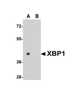
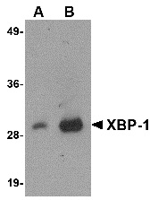
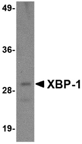
Western Blot Validation in Human HepG2 Cell Lysate. Loading: 15 µg of lysates per lane. Antibodies: XBP-1 orb1240297 (1 µg/mL), 1h incubation at RT in 5% NFDM/TBST. Secondary: Goat anti-rabbit IgG HRP conjugate at 1:10000 dilution. (A) the absence and (B) the presence of blocking peptide.
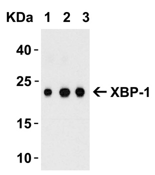
Western Blot Validation with Recombinant Protein. Loading: 100 ng of human XBP-1 recombinant protein per lane. Antibodies: XBP-1 orb1240297 (A: 0.5 µg/mL, B: 1 µg/mL and C: 2 µg/mL), 1h incubation at RT in 5% NFDM/TBST. Secondary: Goat anti-rabbit IgG HRP conjugate at 1:10000 dilution. Observed at around 23kD.
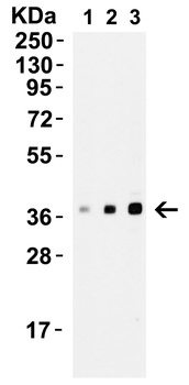
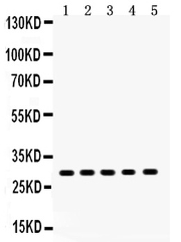
Western Blot Validation of XBP-1 in A549 Cells. Loading: 15 µg of A549 cell lysate. Antibodies: XBP-1 orb1240297, 1h incubation at RT in 5% NFDM/TBST. Secondary: Goat anti-rabbit IgG HRP conjugate at 1:10000 dilution. Lane1: 0.25 µg/mL, Lane2: 0.5 µg/mL, Lane3: 1 µg/mL.
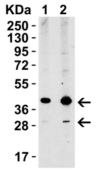
Western Blot Validation of XBP-1 in HepG2 Cells. Loading: 15 µg of HepG2 cell lysate. Antibodies: XBP-1 orb1240297, 1h incubation at RT in 5% NFDM/TBST. Secondary: Goat anti-rabbit IgG HRP conjugate at 1:10000 dilution. Lane1: 1 µg/mL, Lane2: 2 µg/mL.
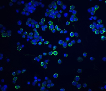
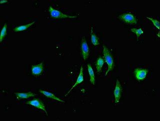
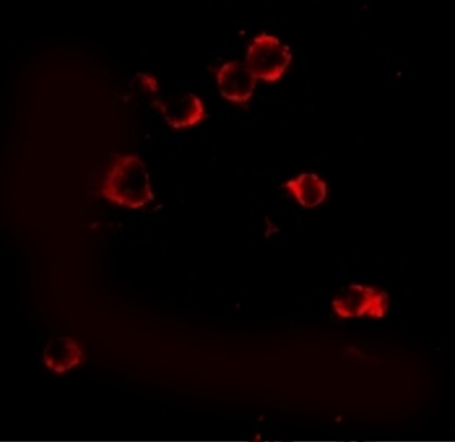
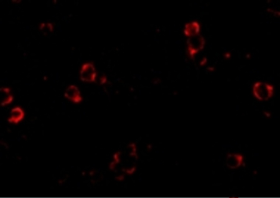
Immunofluorescence Validation of XBP-1 in Human HepG2 Cells. Immunofluorescent analysis of 4% paraformaldehyde-fixed HepG2 cells labeling XBP-1 with orb1240297 at 20 µg/mL, followed by goat anti-rabbit IgG secondary antibody at 1/500 dilution (green) and DAPI staining (blue).
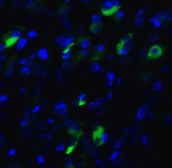
Immunofluorescence Validation of XBP-1 in Human Pancreas Tissue. Immunofluorescent analysis of 4% paraformaldehyde-fixed human pancreas tissue labeling XBP-1 with orb1240297 at 20 µg/mL, followed by goat anti-rabbit IgG secondary antibody at 1/500 dilution (green) and DAPI staining (blue).
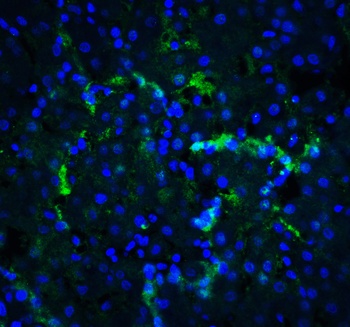
Immunofluorescence Validation of XBP-1 in Human Liver Tissue. Immunofluorescent analysis of 4% paraformaldehyde-fixed Human Liver Tissue labeling XBP-1 with orb1240297 at 20 µg/mL, followed by goat anti-rabbit IgG secondary antibody at 1/500 dilution (green) and DAPI staining (blue).
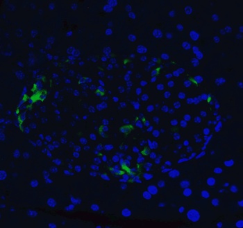
Immunofluorescence Validation of XBP-1 in Mouse Pancreas Tissue. Immunofluorescent analysis of 4% paraformaldehyde-fixed mouse pancreas tissue labeling XBP-1 with orb1240297 at 20 µg/mL, followed by goat anti-rabbit IgG secondary antibody at 1/500 dilution (green) and DAPI staining (blue).
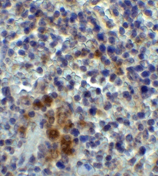
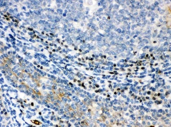
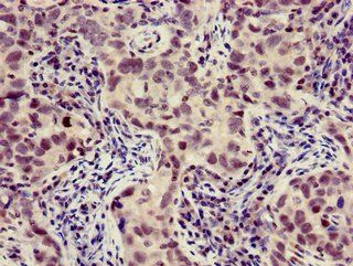
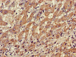
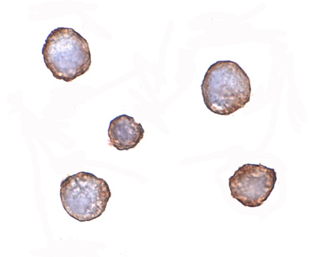
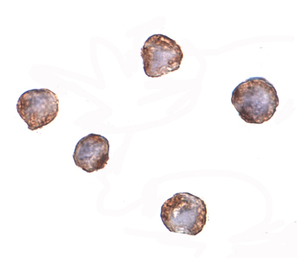
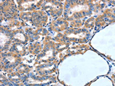
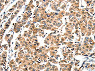
Immunohistochemistry Validation of XBP-1 in Mouse Spleen Tissue. Immunohistochemical analysis of paraffin-embedded mouse spleen tissue using anti-XBP-1 antibody (orb1240297) at 2 µg/ml. Tissue was fixed with formaldehyde and blocked with 10% serum for 1 h at RT; antigen retrieval was by heat mediation with a citrate buffer (pH6). Samples were incubated with primary antibody overnight at 4°C. A goat anti-rabbit IgG H&L (HRP) at 1/250 was used as secondary. Counter stained with Hematoxylin.
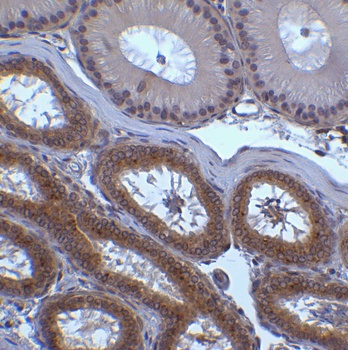
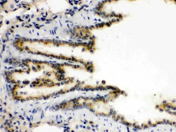
Immunohistochemistry Validation of XBP-1 in Mouse Testis Tissue. Immunohistochemical analysis of paraffin-embedded mouse testis tissue using anti-XBP-1 antibody (orb1240297) at 2 µg/ml. Tissue was fixed with formaldehyde and blocked with 10% serum for 1 h at RT; antigen retrieval was by heat mediation with a citrate buffer (pH6). Samples were incubated with primary antibody overnight at 4 °C. A goat anti-rabbit IgG H&L (HRP) at 1/250 was used as secondary. Counter stained with Hematoxylin.
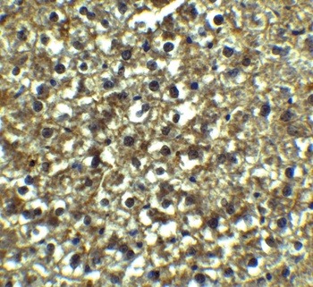
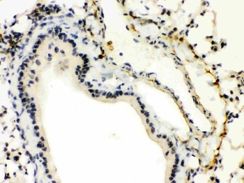
Immunohistochemistry Validation of XBP-1 in Rat Liver Tissue. Immunohistochemical analysis of paraffin-embedded Rat Liver Tissue using anti-XBP-1 antibody (orb1240297) at 5 µg/ml. Tissue was fixed with formaldehyde and blocked with 10% serum for 1 h at RT; antigen retrieval was by heat mediation with a citrate buffer (pH6). Samples were incubated with primary antibody overnight at 4°C. A goat anti-rabbit IgG H&L (HRP) at 1/250 was used as secondary. Counter stained with Hematoxylin.
- Item 1 of 5
XBP1 Antibody [orb259640]
FC, ICC, IF, IHC, WB
Hamster
Human, Mouse, Rat
Rabbit
Polyclonal
Unconjugated
10 μg, 100 μg - Item 1 of 3
- Item 1 of 3
- Item 1 of 3
- Item 1 of 2
Submit a review
Filter by Rating

- 5 stars

- 4 stars
- 3 stars
- 2 stars
- 1 stars