You have no items in your shopping cart.
Cart summary
Item 1 of 4
Item 1 of 4
WASP Antibody
Catalog Number: orb259636
| Catalog Number | orb259636 |
|---|---|
| Category | Antibodies |
| Description | WASP Antibody |
| Species/Host | Rabbit |
| Clonality | Polyclonal |
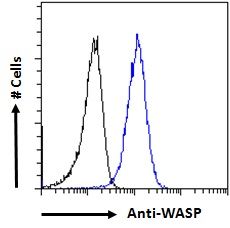
| Tested applications | ICC, IHC, WB |
| Reactivity | Human, Mouse, Rat |
| Isotype | Rabbit IgG |
| Immunogen | A synthetic peptide corresponding to a sequence at the C-terminus of human WASP (129-156aa ADEDEAQAFRALVQEKIQKRNQRQSGDR), different from the related mouse sequence by two amino acids. |
| Concentration | Adding 0.2 ml of distilled water will yield a concentration of 500 μg/ml. |
| Dilution range | Immunocytochemistry , 0.5-1μg/ml, Human, -Immunohistochemistry (Frozen Section), 0.5-1μg/ml, Human, -Immunohistochemistry (Paraffin-embedded Section), 0.5-1μg/ml, Human, By HeatWestern blot, 0.1-0.5μg/ml, Human, Mouse, Rat |
| Form/Appearance | Lyophilized |
| Conjugation | Unconjugated |
| MW | 52913 MW |
| UniProt ID | P42768 |
| Storage | Store at -20˚C for one year from date of receipt. After reconstitution, at 4˚C for one month. It can also be aliquotted and stored frozen at -20˚C for six months. Avoid repeated freeze-thaw cycles. |
| Alternative names | Wiskott-Aldrich syndrome protein;WASp;WAS;IMD2; Read more... |
| Note | For research use only |
| Application notes | WB: The detection limit for WASP is approximately 0.1ng/lane under reducing conditions. Tested Species: In-house tested species with positive results. By Heat: Boiling the paraffin sections in 10mM citrate buffer, pH6.0, for 20mins is required for the staining of formalin/paraffin sections. Other applications have not been tested. Optimal dilutions should be determined by end users. . Add 0.2ml of distilled water will yield a concentration of 500ug/ml. |
| Expiration Date | 12 months from date of receipt. |
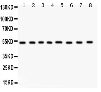
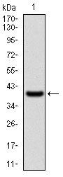
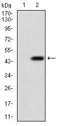
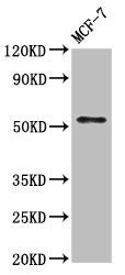
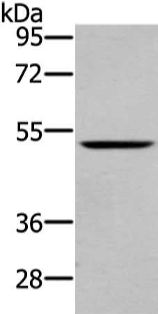
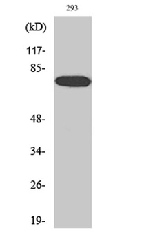
WB analysis of WASP using anti-WASP antibody.Lane 1:Rat Liver tissue;2:Human Placenta tissue;3:Rat Spleen tissue;4:Rat Pancreas tissue;5:HEPG2 cell;6:HELA cell;7;HEPA cell;8;22RV1 Cell.
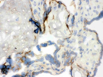
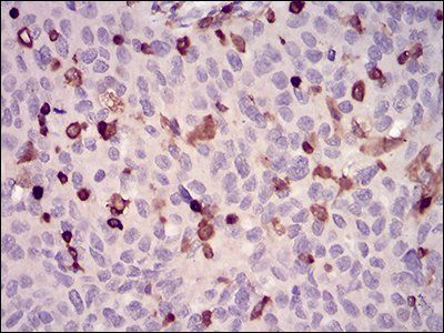
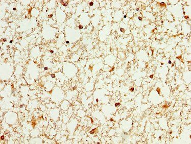
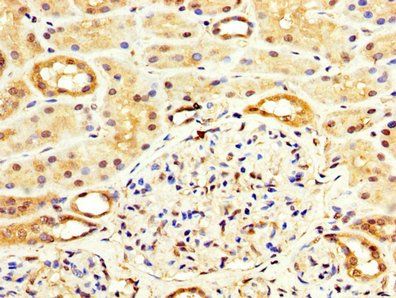
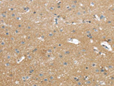
IHC analysis of WAS using anti-WAS antibody.WAS was detected in frozen section of human placenta tissue.
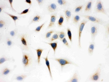
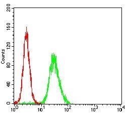
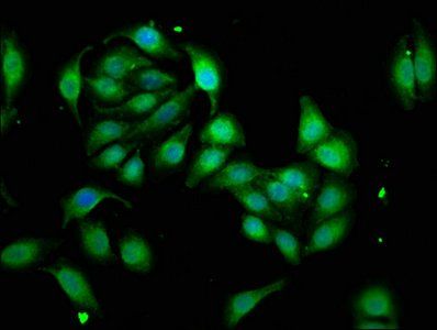
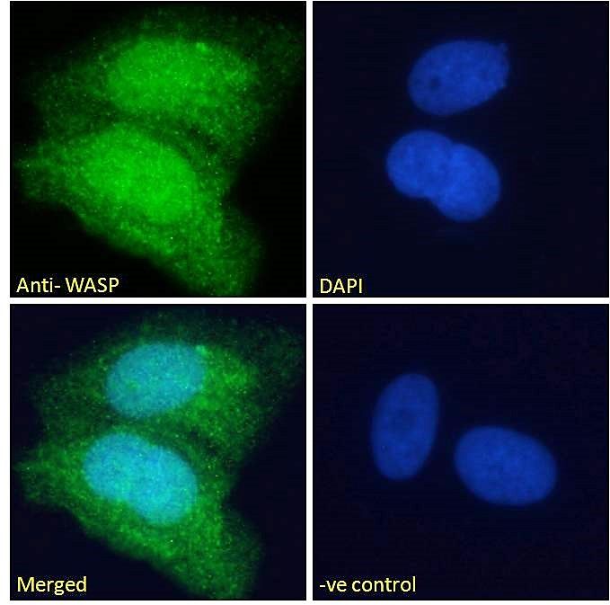
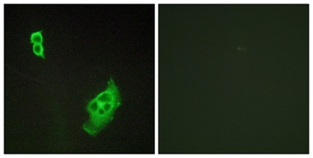
IHC analysis of WAS using anti-WAS antibody.WAS was detected in immunocytochemical section of a549 cell.
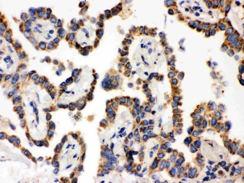
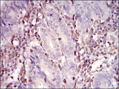
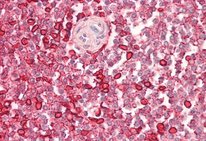
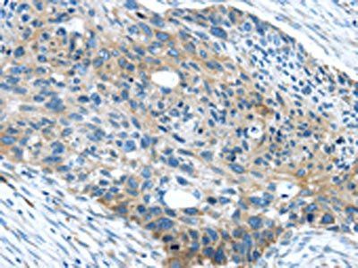
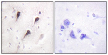
IHC analysis of WAS using anti-WAS antibody.WAS was detected in paraffin-embedded section of human lung cancer tissue.
- Item 1 of 6
- Item 1 of 4
- Item 1 of 4
WAS antibody [orb18966]
ELISA, FC, IF, IHC
Canine, Human, Mouse, Rat
Goat
Polyclonal
Unconjugated
100 μg - Item 1 of 3
- Item 1 of 4
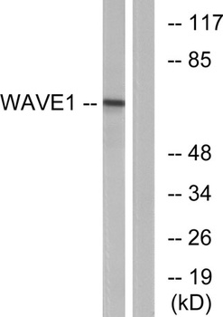
WAVE1 antibody [orb766577]
ELISA, IF, IHC-P, WB
Human, Mouse, Rat
Rabbit
Polyclonal
Unconjugated
50ul, 100ul
Submit a review
Filter by Rating

- 5 stars

- 4 stars
- 3 stars
- 2 stars
- 1 stars