You have no items in your shopping cart.
Cart summary
Item 1 of 4
Item 1 of 4
TPH1 Antibody
Catalog Number: orb1263580

| Catalog Number | orb1263580 |
|---|---|
| Category | Antibodies |
| Description | TPH1 Antibody |
| Species/Host | Rabbit |
| Clonality | Polyclonal |
| Tested applications | FC, IF, IHC-P, WB |
| Predicted Reactivity | Gallus, Rabbit, Rat, Xenopus |
| Reactivity | Human |
| Isotype | Rabbit Ig |
| Immunogen | This TPH1 antibody is generated from rabbits immunized with a KLH conjugated synthetic peptide between 35-62 amino acids from the N-terminal region of human TPH1. |
| Antibody Type | Primary Antibody |
| Concentration | batch dependent |
| Form/Appearance | Liquid |
| Conjugation | Unconjugated |
| MW | 51 kDa |
| Target | TPH1 |
| UniProt ID | P17752 |
| NCBI | P17752 |
| Storage | Maintain refrigerated at 2-8°C for up to 2 weeks. For long term storage store at -20°C in small aliquots to prevent freeze-thaw cycles. |
| Buffer/Preservatives | Supplied in PBS with 0.09% (W/V) sodium azide. |
| Alternative names | Tryptophan 5-hydroxylase 1, Tryptophan 5-monooxyge Read more... |
| Note | For research use only |
| Application notes | For WB starting dilution is: 1:500For IHC-P starting dilution is: 1:50~100For FACS starting dilution is: 1:10~50For IF starting dilution is: 1:10~50 |
| Expiration Date | 12 months from date of receipt. |
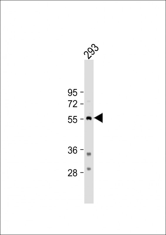
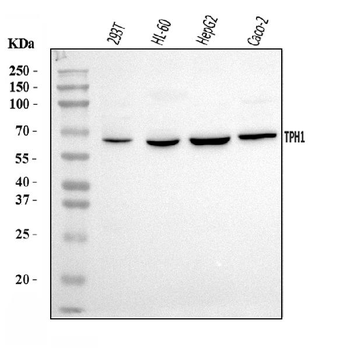
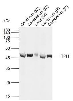
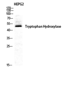
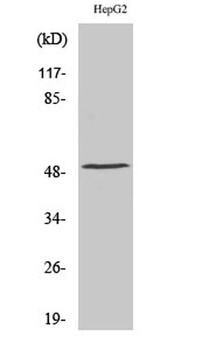
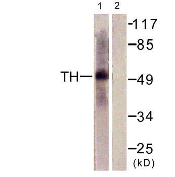
Western Blot at 1:500 dilution + 293 whole cell lysate Lysates/proteins at 20 ug per lane.
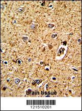
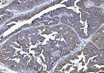
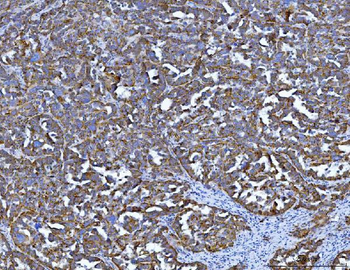
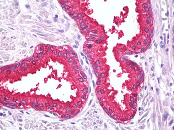
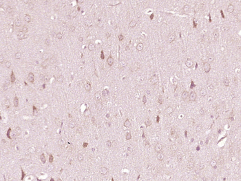
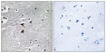
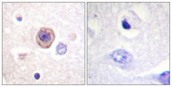
Formalin-fixed and paraffin-embedded human brain tissue with TPH1 Antibody (N-term), which was peroxidase-conjugated to the secondary antibody, followed by DAB staining.
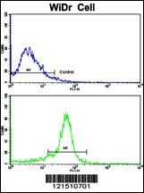
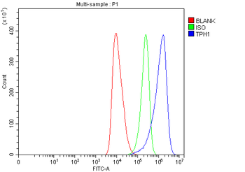
Flow cytometric analysis of widr cells using TPH1 Antibody (N-term) (bottom histogram) compared to a negative control cell (top histogram). FITC-conjugated goat-anti-rabbit secondary antibodies were used for the analysis.
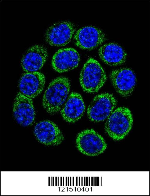
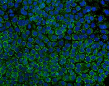
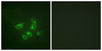
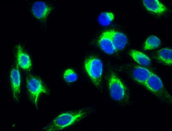
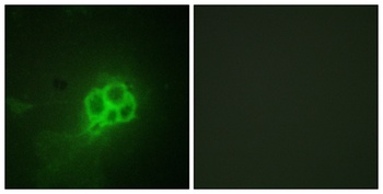
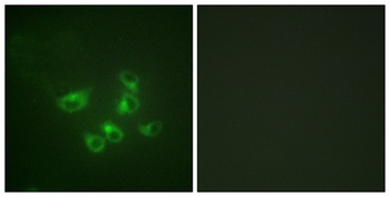
Confocal immunofluorescent analysis of TPH1 Antibody with 293 cell followed by Alexa Fluor 488-conjugated goat anti-rabbit lgG (green). DAPI was used to stain the cell nuclear (blue).
- Item 1 of 6
Anti-Tryptophan Hydroxylase/TPH1 Antibody [orb1098051]
ELISA, FC, ICC, IF, IHC, WB
Human
Rabbit
Polyclonal
Unconjugated
10 μg, 100 μg - Item 1 of 3
TPH1/Tryptophan Hydroxylase Antibody [orb1538880]
ELISA, IF, IHC, IHC-P, WB
Human, Mouse, Rat
Rabbit
Polyclonal
Unconjugated
50 μl - Item 1 of 2
TPH Rabbit Polyclonal Antibody [orb11500]
IF, IHC-Fr, IHC-P, WB
Canine, Equine, Gallus, Human, Rabbit
Mouse, Rat
Rabbit
Polyclonal
Unconjugated
100 μl, 200 μl, 50 μl - Item 1 of 4
TPH1 rabbit pAb [orb770319]
ELISA, IF, IHC-P, WB
Human, Mouse, Rat
Polyclonal
Unconjugated
100 μl, 50 μl - Item 1 of 4
TPH1 rabbit pAb [orb766505]
ELISA, IF, IHC-P, WB
Human, Mouse, Rat
Polyclonal
Unconjugated
50 μl, 100 μl