You have no items in your shopping cart.
Cart summary
Item 1 of 3
Item 1 of 3
TP53INP1 Antibody
Catalog Number: orb1239300
| Catalog Number | orb1239300 |
|---|---|
| Category | Antibodies |
| Description | TP53INP1 Antibody |
| Species/Host | Rabbit |
| Clonality | Polyclonal |
| Tested applications | ELISA, IF, IHC-P, WB |
| Reactivity | Human, Mouse, Rat |
| Isotype | IgG |
| Immunogen | p53DINP1 antibody was raised with a synthetic peptide corresponding to 14 amino acids near the amino terminus of human p53DINP1.The immunogen is located within the first 50 amino acids of p53DINP1. |
| Concentration | 1 mg/mL |
| Dilution range | p53DINP1 antibody can be used for detection of p53DINP1 by Western blot at 0.5 - 1 μg/mL. Antibody can also be used for immunohistochemistry starting at 2 μg/mL. For immunofluorescence start at 20 μg/mL.Antibody validated: Western Blot in human samples; Immunohistochemistry in mouse samples and Immunofluorescence in human samples. All other applications and species not yet tested. |
| Form/Appearance | Liquid |
| Conjugation | Unconjugated |
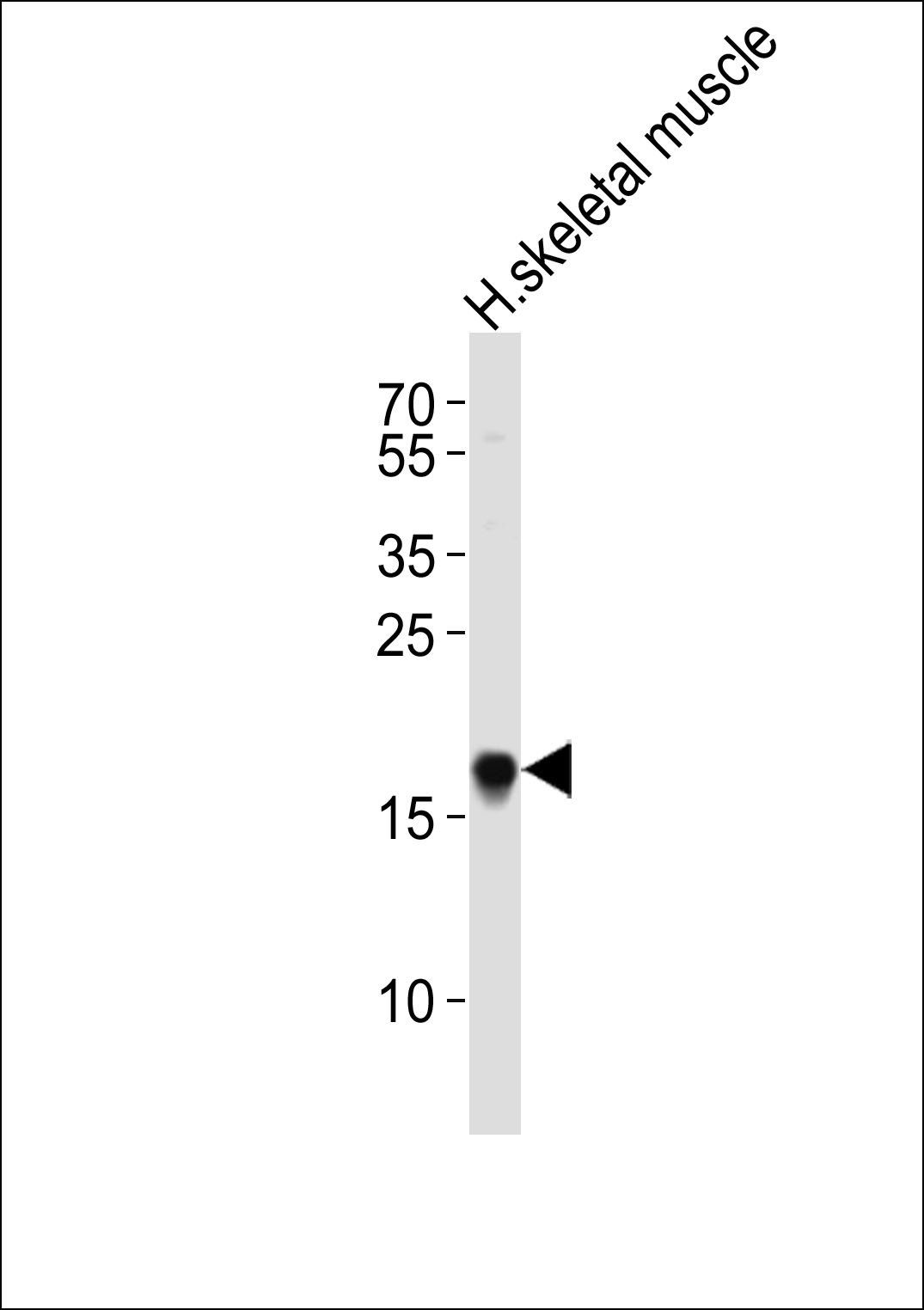
| MW | Predicted: 18, 27 kDa Observed: 30 kDa |
| Target | TP53INP1 |
| UniProt ID | Q96A56 |
| NCBI | Q96A56 |
| Storage | p53DINP1 antibody can be stored at 4°C for three months and -20°C, stable for up to one year. As with all antibodies care should be taken to avoid repeated freeze thaw cycles. Antibodies should not be exposed to prolonged high temperatures. |
| Buffer/Preservatives | p53DINP1 Antibody is supplied in PBS containing 0.02% sodium azide. |
| Alternative names | p53DINP1 Antibody: SIP, Teap, p53DINP1, TP53DINP1, Read more... |
| Note | For research use only |
| Application notes | p53DINP1 antibody can be used for detection of p53DINP1 by Western blot at 0.5 - 1 μg/mL. Antibody can also be used for immunohistochemistry starting at 2 μg/mL. For immunofluorescence start at 20 μg/mL.Antibody validated: Western Blot in human samples; Immunohistochemistry in mouse samples and Immunofluorescence in human samples. All other applications and species not yet tested. |
| Expiration Date | 12 months from date of receipt. |
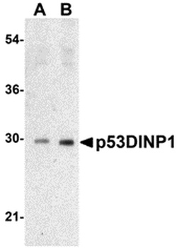
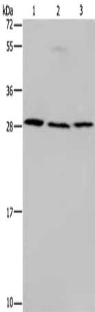
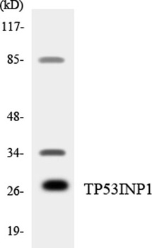
Western blot analysis of p53DINP1 expression in human lung tissue lysate with p53DINP1 antibody at (A) 0.5 and (B) 1 µg/mL.
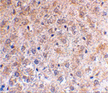
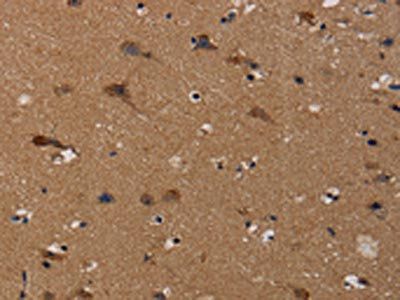
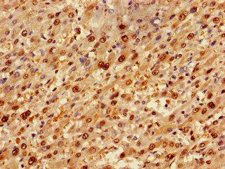
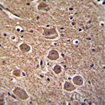
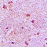
Immunohistochemical staining of mouse liver using p53DINP1 antibody at 2 µg/mL.
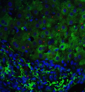
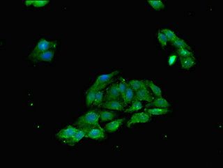
Immunofluorescence of p53DINP1 in human liver tissue with p53DINP1 antibody at 5 µg/ml. Green: p53DINP1 antibody (orb1239300) Red: Phylloidin staining Blue: DAPI staining.
- Item 1 of 3
- Item 1 of 4
- Item 1 of 2
- Item 1 of 3
- Item 1 of 2
TP53INP1 antibody [orb215345]
IH, WB
Human, Primate
Rabbit
Polyclonal
Unconjugated
200 μl, 100 μl, 30 μl
Submit a review
Filter by Rating

- 5 stars

- 4 stars
- 3 stars
- 2 stars
- 1 stars