You have no items in your shopping cart.
Cart summary
Item 1 of 4
Item 1 of 4
Tenascin antibody
Catalog Number: orb44503

| Catalog Number | orb44503 |
|---|---|
| Category | Antibodies |
| Description | Mouse Monoclonal to Tenascin. |
| Clonality | Monoclonal |
| Clone Number | T2H5 |
| Tested applications | IHC-P, IP, WB |
| Reactivity | Human |
| Isotype | Mouse IgG1 |
| Immunogen | Protein preparation from a homogenate of a human mammary tumour specimen. |
| Concentration | 1 mg/ml |
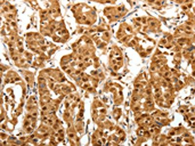
| Dilution range | Immunohistochemistry (paraffin sections): Immunohistochemical detection of tenascin is valuable for studies of tissue differentiation and tumour growth. The antibody T2H5 is excellent for staining of paraffin-embedded tissue sections.Western blotting: Recommended dilution: 1-2 μg/ml. |
| Purity | Purified by protein-A affinity chromatography. |
| Conjugation | Unconjugated |
| Target | Tenascin C |
| Entrez | 3371 |
| UniProt ID | P24821 |
| Storage | Store at 2-8°C. Do not freeze. |
| Buffer/Preservatives | Tris buffered saline (TBS), pH 8.0, 15 mM sodium azide |
| Alternative names | Tenascin Read more... |
| Note | For research use only |
| Application notes | Immunohistochemistry (paraffin sections): Immunohistochemical detection of tenascin is valuable for studies of tissue differentiation and tumour growth. The antibody T2H5 is excellent for staining of paraffin-embedded tissue sections. Heat-mediated antigen retrieval in citrate buffer.Western blotting: Recommended dilution: 1-2 μg/ml. |
| Expiration Date | 12 months from date of receipt. |
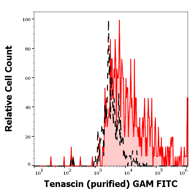
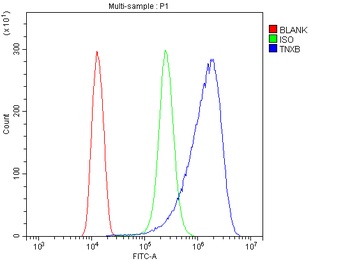
Separation of U-87 MG cells stained using anti-tenascin C (T2H5) purified antibody (concentration in sample 12 µg/ml, GAM FITC, red-filled) from U-87 MG cells unstained by primary antibody (GAM FITC, black-dashed) in flow cytometry analysis (surface staining).
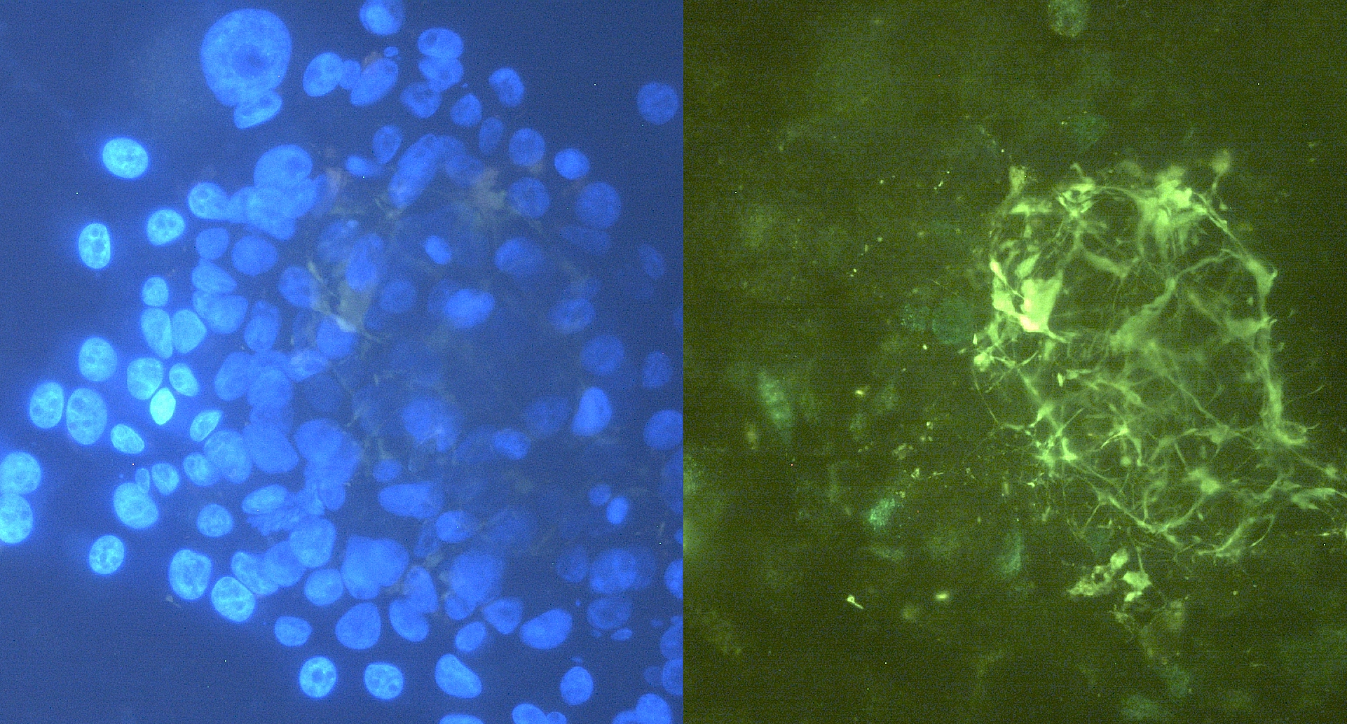
Immunocytochemistry staining of tenascin C in U-87 MG cells using purified mouse monoclonal antibody T2H7 (concentration in sample 12 µg/ml, GAM FITC, right picture) vs. Hoechst 34580 nuclear staining (left picture).
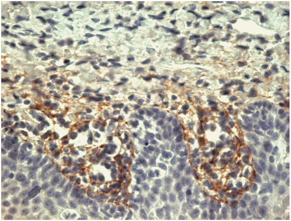
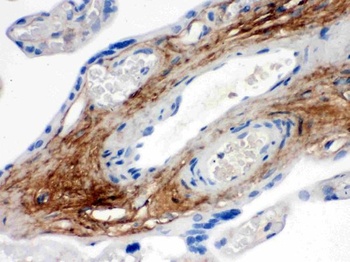
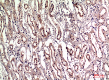
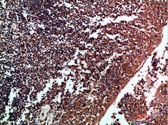
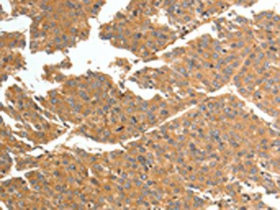
Immunohistochemistry staining of tonsil (paraffin-embedded sections) with anti-human tenascin C (T2H5).
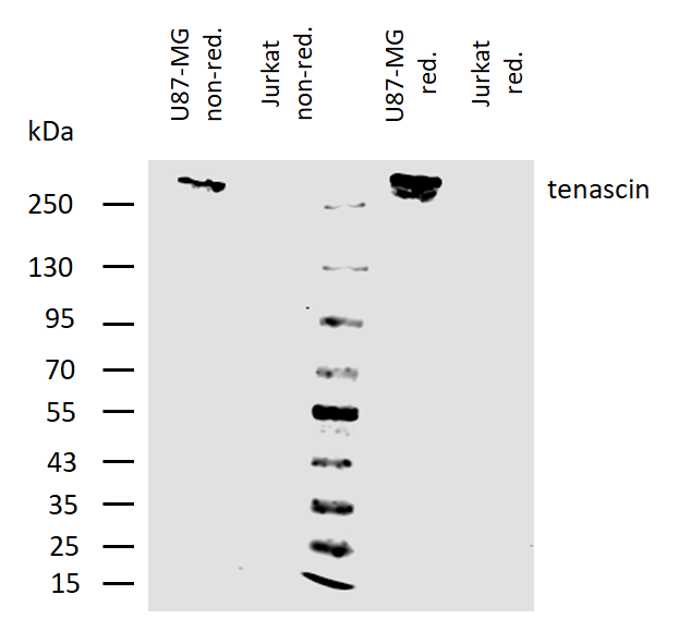
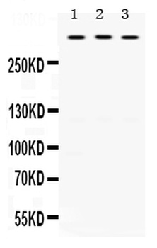
Western blotting analysis of human tenascin C using mouse monoclonal antibody T2H5 on lysates of U87-MG cell line and Jurkat cell line (tenascin non-expressing cell line; negative control) under non-reducing and reducing conditions. Cells were lysed by 50 mM TRIS-Cl pH 6.6, 4M urea, 4% SDS, samples were mixed and heated (100°C, 5 min) with reducing and non-reducing SDS-loading buffer, then resolved using 7.5% Tris-glycine SDS gel electrophoresis. Nitrocellulose membrane was probed with 2 µg/ml of mouse anti-tenascin monoclonal antibody followed by IRDye800-conjugated anti-mouse secondary antibody. Tenascin C was detected slightly above 250 kDa.
- Item 1 of 4
- Item 1 of 4
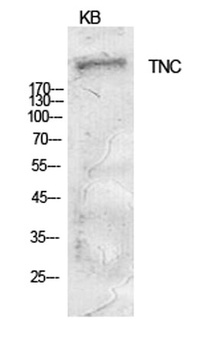
Tenascin-C antibody [orb767153]
ELISA, IHC-P, WB
Human, Mouse, Rat
Rabbit
Polyclonal
Unconjugated
100ul, 50ul - Item 1 of 2
- Item 1 of 2
- Item 1 of 2

Submit a review
Filter by Rating

- 5 stars

- 4 stars
- 3 stars
- 2 stars
- 1 stars
