You have no items in your shopping cart.
Cart summary
Item 1 of 5
Item 1 of 5
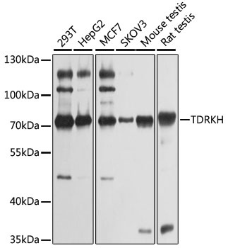
TDRKH Antibody
Catalog Number: orb2635202

| Catalog Number | orb2635202 |
|---|---|
| Category | Antibodies |
| Description | Participates in the primary piRNA biogenesis pathway and is required during spermatogenesis to repress transposable elements and prevent their mobilization, which is essential for the germline integrity. The piRNA metabolic process mediates the repression of transposable elements during meiosis by forming complexes composed of piRNAs and Piwi proteins and govern the methylation and subsequent repression of transposons. Required for the final steps of primary piRNA biogenesis by participating in the processing of 31-37 nt intermediates into mature piRNAs. May act in pi-bodies and piP-bodies by transferring piRNA precursors or intermediates to or between these granules. |
| Clonality | Monoclonal |
| Species/Host | Mouse |
| Isotype | Mouse IgG2b |
| Conjugation | Unconjugated |
| Reactivity | Human |
| Immunogen | Recombinant full-length human TDRKH protein was used as the immunogen for the TDRKH antibody. |
| UniProt ID | Q9Y2W6 |
| Tested applications | FACS, IF, IHC-P, WB |
| Dilution range | Flow cytometry: 1-2ug/million cells,Immunofluorescence: 1-2ug/ml,Western blot: 1-2ug/ml,Immunohistochemistry (FFPE): 1-2ug/ml |
| Application notes | Optimal dilution of the TDRKH antibody should be determined by the researcher. |
| Antibody Type | Primary Antibody |
| Clone Number | PCRP-TDRKH-1H2 |
| Formula | 0.2 mg/ml in 1X PBS with 0.1 mg/ml BSA (US sourced), 0.05% sodium azide |
| Storage | Maintain refrigerated at 2-8°C for up to 2 weeks. For long term storage store at -20°C in small aliquots to prevent freeze-thaw cycles. |
| Hazard Information | This TDRKH antibody is available for research use only. |
| Note | For research use only |
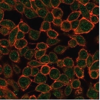
Immunofluorescent staining of PFA-fixed human HeLa cells using TDRKH antibody (green, clone PCRP-TDRKH-1H2) and phalloidin (red).
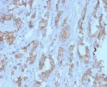
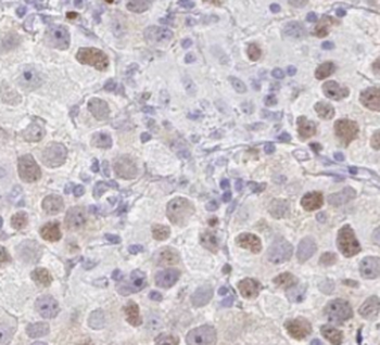
IHC staining of FFPE human ovarian carcinoma tissue with TDRKH antibody (clone PCRP-TDRKH-1H2) at 2 ug/ml. HIER: boil tissue sections in pH9 10mM Tris with 1mM EDTA for 20 min and allow to cool before testing.
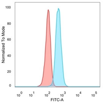
FACS staining of PFA-fixed human HeLa cells with TDRKH antibody (blue, clone PCRP-TDRKH-1H2), and unstained cells (red).
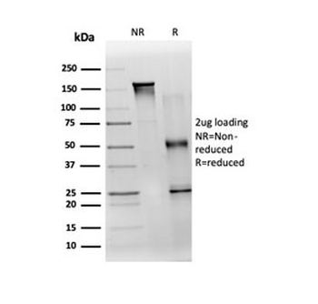
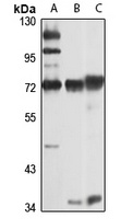
SDS-PAGE analysis of purified, BSA-free TDRKH antibody (clone PCRP-TDRKH-1H2) as confirmation of integrity and purity.
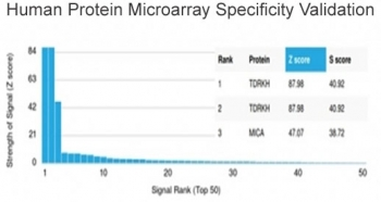
Analysis of HuProt (TM) microarray containing more than 19000 full-length human proteins using TDRKH antibody (clone PCRP-TDRKH-1H2). These results demonstrate the foremost specificity of the PCRP-TDRKH-1H2 mAb. Z- and S- score: The Z-score represents the strength of a signal that an antibody (in combination with a fluorescently-tagged anti-IgG secondary Ab) produces when binding to a particular protein on the HuProt (TM) array. Z-scores are described in units of standard deviations (SD's) above the mean value of all signals generated on that array. If the targets on the HuProt (TM) are arranged in descending order of the Z-score, the S-score is the difference (also in units of SD's) between the Z-scores. The S-score therefore represents the relative target specificity of an Ab to its intended target.
- Item 1 of 5
- Item 1 of 3
TDRKH Antibody [orb395667]
ELISA, IF, IHC, IP, WB
Human, Mouse, Rat
Rabbit
Polyclonal
Unconjugated
100 μg, 50 μg - Item 1 of 1
Anti-TDRKH Antibody [orb668385]
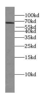
WB
Human, Mouse, Rat
Rabbit
Polyclonal
Unconjugated
50 μl, 100 μl, 200 μl 
- Item 1 of 1