You have no items in your shopping cart.
Cart summary
Item 1 of 2
Item 1 of 2
Synaptophysin SYP Rabbit Monoclonal Antibody
Catalog Number: orb548345
| Catalog Number | orb548345 |
|---|---|
| Category | Antibodies |
| Description | Synaptophysin SYP Rabbit Monoclonal Antibody |
| Species/Host | Rabbit |
| Clonality | Monoclonal |
| Clone Number | BGB-19 |
| Tested applications | ICC, IF, IHC, WB |
| Reactivity | Human, Mouse, Rat |
| Isotype | Rabbit IgG |
| Immunogen | A synthesized peptide derived from human Synaptophysin |
| Concentration | Actual concentration vary by lot. Use suggested dilution ratio to decide dilution procedure. |
| Dilution range | WB 1:5000-1:10000IHC 1:50-1:200ICC/IF 1:50-1:200 |
| Form/Appearance | Liquid |
| Conjugation | Unconjugated |
| MW | 33845 MW |
| UniProt ID | P08247 |
| Storage | Store at -20°C for one year. For short term storage and frequent use, store at 4°C for up to one month. Avoid repeated freeze-thaw cycles. |
| Alternative names | Synaptophysin;Major synaptic vesicle protein p38;S Read more... |
| Note | For research use only |
| Expiration Date | 12 months from date of receipt. |
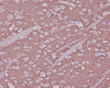
Immunohistochemical analysis of paraffin-embedded mouse brain, using Synaptophysin Antibody (orb548345) SYP was detected in paraffin-embedded tissue section. Heat mediated antigen retrieval was performed in citrate buffer (pH6, epitope retrieval solution) for 20 mins. The tissue section was blocked with 10% goat serum. The tissue section was then incubated with 1ug/ml rabbit anti-SYP Antibody (orb548345) overnight at 4°C. Biotinylated goat anti-rabbit IgG was used as secondary antibody and incubated for 30 minutes at 37°C. The tissue section was developed using Strepavidin-Biotin-Complex (SABC) (Catalog # orb90444) with DAB as the chromogen.
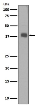
Western blot analysis of Synaptophysin expression in SH-SY5Y cell lysate (orb548345). Electrophoresis was performed on a 5-20% SDS-PAGE gel at 70V (Stacking gel)/90V (Resolving gel) for 2-3 hours. The sample well of each lane was loaded with 50ug of sample under reducing conditions. After Electrophoresis, proteins were transferred to a Nitrocellulose membrane at 150mA for 50-90 minutes. Blocked the membrane with 5% Non-fat Milk/TBS for 1.5 hour at RT. The membrane was incubated with rabbit anti-SYP monoclonal antibody (Catalog # orb548345) overnight at 4°C, then washed with TBS-0.1%Tween 3 times with 5 minutes each and probed with a goat anti-rabbit IgG-HRP secondary antibody at a dilution of 1:10000 for 1.5 hour at RT. The signal is developed using an Enhanced Chemiluminescent detection (ECL) kit (Catalog # orb90503) with Tanon 5200 system. A specific band was detected for SYP.
- Item 1 of 2
Synaptophysin SYP Rabbit Monoclonal Antibody [orb548344]
FC, ICC, IF, IHC, IP, WB
Human, Mouse, Rat
Rabbit
Monoclonal
Unconjugated
30 μl, 100 μl 
Synaptophysin (Neuroendocrine Marker) antibody [orb1410438]
IHC-P
Human
Rabbit
Monoclonal
Unconjugated
20 μg, 100 μg
Submit a review
Filter by Rating

- 5 stars

- 4 stars
- 3 stars
- 2 stars
- 1 stars