You have no items in your shopping cart.
Cart summary
Item 1 of 8
Item 1 of 8
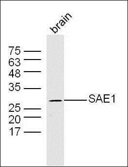
Sumo 1/SUMO1 Antibody
Catalog Number: orb692211
| Catalog Number | orb692211 |
|---|---|
| Category | Antibodies |
| Description | Sumo 1/SUMO1 Antibody |
| Species/Host | Rabbit |
| Clonality | Polyclonal |
| Tested applications | FC, ICC, IF, IHC |
| Reactivity | Human, Mouse, Rat |
| Isotype | Rabbit IgG |
| Immunogen | A synthetic peptide corresponding to a sequence of human Sumo 1/SUMO1 (HLKKLKESYCQRQGVPMNSLRFLFEGQRIADNHTPKEL). |
| Concentration | Adding 0.2 ml of distilled water will yield a concentration of 500 μg/ml. |
| Dilution range | Immunohistochemistry (Paraffin-embedded Section), 1-2μg/ml, Human, Mouse, Rat Immunocytochemistry/Immunofluorescence, 5μg/ml, Human Flow Cytometry, 1-3μg/1x106 cells, Human, Mouse |
| Form/Appearance | Lyophilized |
| Conjugation | Unconjugated |
| MW | 12 kDa |
| UniProt ID | P63165 |
| Storage | Store at -20˚C for one year from date of receipt. After reconstitution, at 4˚C for one month. It can also be aliquotted and stored frozen at -20˚C for six months. Avoid repeated freeze-thaw cycles. |
| Note | For research use only |
| Application notes | Tested Species: In-house tested species with positive results. Other applications have not been tested. Optimal dilutions should be determined by end users. Add 0.2ml of distilled water will yield a concentration of 500μg/ml. |
| Expiration Date | 12 months from date of receipt. |
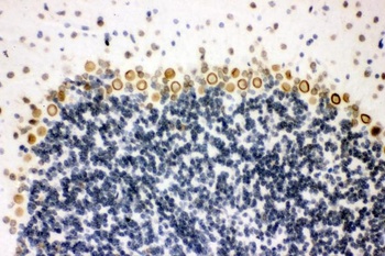
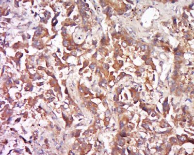
IHC analysis of Sumo 1/SUMO1 using anti-Sumo 1/SUMO1 antibody (orb692211). Sumo 1/SUMO1 was detected in paraffin-embedded section of mouse brain tissue. Heat mediated antigen retrieval was performed in EDTA buffer (pH8.0, epitope retrieval solution). The tissue section was blocked with 10% goat serum. The tissue section was then incubated with 2μg/ml rabbit anti-Sumo 1/SUMO1 Antibody (orb692211) overnight at 4°C. Biotinylated goat anti-rabbit IgG was used as secondary antibody and incubated for 30 minutes at 37°C. The tissue section was developed using Strepavidin-Biotin-Complex (SABC) (Catalog # orb90444) with DAB as the chromogen.
IHC analysis of Sumo 1/SUMO1 using anti-Sumo 1/SUMO1 antibody (orb692211). Sumo 1/SUMO1 was detected in paraffin-embedded section of mouse brain tissue. Heat mediated antigen retrieval was performed in EDTA buffer (pH8.0, epitope retrieval solution). The tissue section was blocked with 10% goat serum. The tissue section was then incubated with 2μg/ml rabbit anti-Sumo 1/SUMO1 Antibody (orb692211) overnight at 4°C. Biotinylated goat anti-rabbit IgG was used as secondary antibody and incubated for 30 minutes at 37°C. The tissue section was developed using Strepavidin-Biotin-Complex (SABC) (Catalog # orb90444) with DAB as the chromogen.
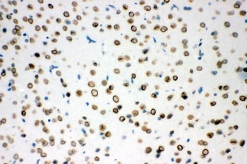
IHC analysis of Sumo 1/SUMO1 using anti-Sumo 1/SUMO1 antibody (orb692211). Sumo 1/SUMO1 was detected in paraffin-embedded section of rat brain tissue. Heat mediated antigen retrieval was performed in EDTA buffer (pH8.0, epitope retrieval solution). The tissue section was blocked with 10% goat serum. The tissue section was then incubated with 2μg/ml rabbit anti-Sumo 1/SUMO1 Antibody (orb692211) overnight at 4°C. Biotinylated goat anti-rabbit IgG was used as secondary antibody and incubated for 30 minutes at 37°C. The tissue section was developed using Strepavidin-Biotin-Complex (SABC) (Catalog # orb90444) with DAB as the chromogen.
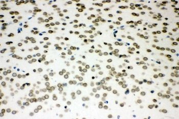
IHC analysis of Sumo 1/SUMO1 using anti-Sumo 1/SUMO1 antibody (orb692211). Sumo 1/SUMO1 was detected in paraffin-embedded section of human intestinal cancer tissue. Heat mediated antigen retrieval was performed in EDTA buffer (pH8.0, epitope retrieval solution). The tissue section was blocked with 10% goat serum. The tissue section was then incubated with 2μg/ml rabbit anti-Sumo 1/SUMO1 Antibody (orb692211) overnight at 4°C. Biotinylated goat anti-rabbit IgG was used as secondary antibody and incubated for 30 minutes at 37°C. The tissue section was developed using Strepavidin-Biotin-Complex (SABC) (Catalog # orb90444) with DAB as the chromogen.
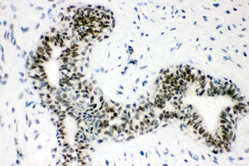
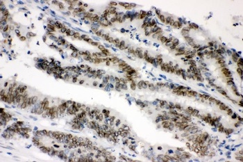
IHC analysis of Sumo 1/SUMO1 using anti-Sumo 1/SUMO1 antibody (orb692211). Sumo 1/SUMO1 was detected in paraffin-embedded section of human mammary cancer tissue. Heat mediated antigen retrieval was performed in EDTA buffer (pH8.0, epitope retrieval solution). The tissue section was blocked with 10% goat serum. The tissue section was then incubated with 2μg/ml rabbit anti-Sumo 1/SUMO1 Antibody (orb692211) overnight at 4°C. Biotinylated goat anti-rabbit IgG was used as secondary antibody and incubated for 30 minutes at 37°C. The tissue section was developed using Strepavidin-Biotin-Complex (SABC) (Catalog # orb90444) with DAB as the chromogen.
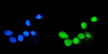
IF analysis of Sumo 1/SUMO1 using anti-Sumo 1/SUMO1 antibody (orb692211). Sumo 1/SUMO1 was detected in immunocytochemical section of A431 cells. Enzyme antigen retrieval was performed using IHC enzyme antigen retrieval reagent (orb90553) for 15 mins. The cells were blocked with 10% goat serum. And then incubated with 5μg/mL rabbit anti-Sumo 1/SUMO1 Antibody (orb692211) overnight at 4°C. DyLight®488 Conjugated Goat Anti-Rabbit IgG was used as secondary antibody at 1:100 dilution and incubated for 30 minutes at 37°C. The section was counterstained with DAPI. Visualize using a fluorescence microscope and filter sets appropriate for the label used.
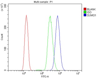
Flow Cytometry analysis of HL-60 cells using anti-Sumo 1/SUMO1 antibody (orb692211). Overlay histogram showing HL-60 cells stained with orb692211 (Blue line). The cells were blocked with 10% normal goat serum. And then incubated with rabbit anti-Sumo 1/SUMO1 Antibody (orb692211, 1μg/1x10^6 cells) for 30 min at 20°C. DyLight®488 conjugated goat anti-rabbit IgG (5-10μg/1x10^6 cells) was used as secondary antibody for 30 minutes at 20°C. Isotype control antibody (Green line) was rabbit IgG (1μg/1x10^6) used under the same conditions. Unlabelled sample (Red line) was also used as a control.
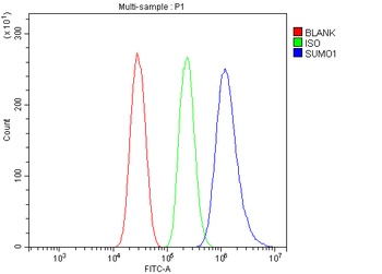
Flow Cytometry analysis of HEPA1-6 cells using anti-Sumo 1/SUMO1 antibody (orb692211). Overlay histogram showing HEPA1-6 cells stained with orb692211 (Blue line). The cells were blocked with 10% normal goat serum. And then incubated with rabbit anti-Sumo 1/SUMO1 Antibody (orb692211, 1 μg/1x10^6 cells) for 30 min at 20°C. DyLight®488 conjugated goat anti-rabbit IgG (5-10 μg/1x10^6 cells) was used as secondary antibody for 30 minutes at 20°C. Isotype control antibody (Green line) was rabbit IgG (1 μg/1x10^6) used under the same conditions. Unlabelled sample (Red line) was also used as a control.
- Item 1 of 2

- Item 1 of 1
- Item 1 of 1
- Item 1 of 1
Submit a review
Filter by Rating

- 5 stars

- 4 stars
- 3 stars
- 2 stars
- 1 stars