You have no items in your shopping cart.
Cart summary
Item 1 of 6
Item 1 of 6
STING1 Antibody / TMEM173 / ERIS / MITA
Catalog Number: orb1823308

| Catalog Number | orb1823308 |
|---|---|
| Category | Antibodies |
| Description | TMEM173 (transmembrane protein 173), also called MITA (Mediator of IRF3 activation), STING1 and ERIS, is a 379 amino acid protein encoded by a gene mapping to human chromosome 5. With 181 million base pairs encoding around 1,000 genes, chromosome 5 is about 6% of human genomic DNA. It is associated with Cockayne syndrome through the ERCC8 gene and familial adenomatous polyposis through the adenomatous polyposis coli (APC) tumor suppressor gene. Treacher Collins syndrome is also chromosome 5 associated and is caused by insertions or deletions within the TCOF1 gene. Deletion of the p arm of chromosome 5 leads to Cri du chat syndrome. Deletion of 5q or chromosome 5 altogether is common in therapy-related acute myelogenous leukemias and myelodysplastic syndrome. |
| Clonality | Recombinant |
| Species/Host | Rabbit |
| Isotype | Rabbit IgG, kappa |
| Conjugation | Unconjugated |
| Reactivity | Human |
| Immunogen | A recombinant partial protein sequence (within amino acids 190-290) from the human protein was used as the immunogen for the MITA antibody. |
| UniProt ID | Q86WV6 |
| Tested applications | IHC-P |
| Dilution range | Immunohistochemistry (FFPE): 1-2ug/ml for 30 min at RT |
| Antibody Type | Primary Antibody |
| Clone Number | STING1/8129R |
| Formula | 0.2 mg/ml in 1X PBS with 0.1 mg/ml BSA (US sourced), 0.05% sodium azide |
| Storage | Aliquot the MITA antibody and store frozen at -20°C or colder. Avoid repeated freeze-thaw cycles. |
| Hazard Information | This MITA antibody is available for research use only. |
| Note | For research use only |
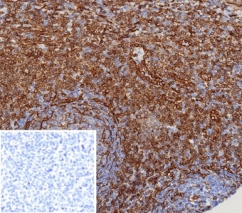
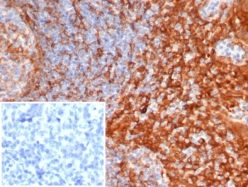
IHC staining of FFPE human tonsil tissue with MITA antibody (clone STING1/8129R). Inset: PBS used in place of primary Ab (secondary Ab negative control). HIER: boil tissue sections in pH9 10 mM Tris with 1 mM EDTA for 20 min and allow to cool before testing.

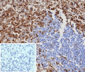
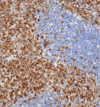
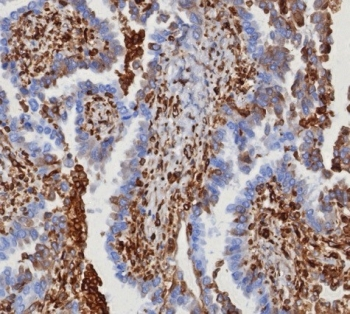
IHC staining of FFPE human spleen tissue with MITA antibody (clone STING1/8129R). HIER: boil tissue sections in pH9 10 mM Tris with 1 mM EDTA for 20 min and allow to cool before testing.
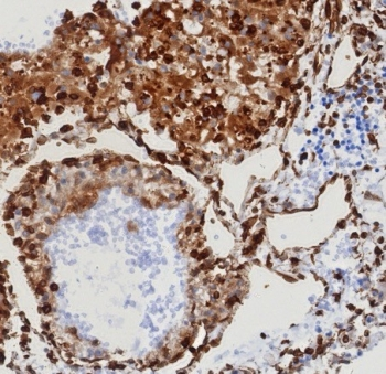
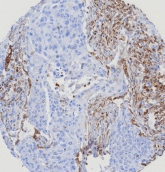
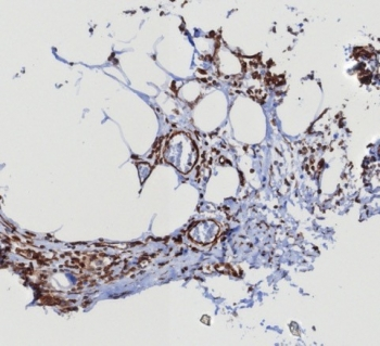
IHC staining of FFPE human serous ovarian carcinoma with MITA antibody (clone STING1/8129R). HIER: boil tissue sections in pH9 10 mM Tris with 1 mM EDTA for 20 min and allow to cool before testing.

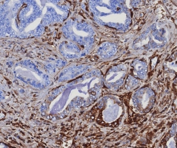
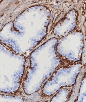
IHC staining of FFPE human prostate carcinoma tissue with MITA antibody (clone STING1/8129R). HIER: boil tissue sections in pH9 10 mM Tris with 1 mM EDTA for 20 min and allow to cool before testing.
IHC staining of FFPE human mammary cancer with MITA antibody (clone STING1/8129R). HIER: boil tissue sections in pH9 10 mM Tris with 1 mM EDTA for 20 min and allow to cool before testing.

IHC staining of FFPE human renal cell carcinoma with MITA antibody (clone STING1/8129R). HIER: boil tissue sections in pH9 10 mM Tris with 1 mM EDTA for 20 min and allow to cool before testing.
- Item 1 of 6
- Item 1 of 6
- Item 1 of 5
STING1 Antibody / TMEM173 / ERIS / MITA [orb1823309]
IHC-P, WB
Human
Mouse
Monoclonal
Unconjugated
100 μg - Item 1 of 5
STING1 Antibody / TMEM173 / ERIS / MITA [orb1823310]
IHC-P, WB
Human
Mouse
Monoclonal
Unconjugated
20 μg - Item 1 of 5
STING1 Antibody / TMEM173 / ERIS / MITA [orb1823311]
IHC-P, WB
Human
Mouse
Monoclonal
Unconjugated
100 μg