You have no items in your shopping cart.
Cart summary
Item 1 of 8
Item 1 of 8
SMARCAD1 Antibody
Catalog Number: orb546363
| Catalog Number | orb546363 |
|---|---|
| Category | Antibodies |
| Description | SMARCAD1 Antibody |
| Species/Host | Rabbit |
| Clonality | Polyclonal |
| Tested applications | ELISA, FC, ICC, IF, IHC, WB |
| Reactivity | Human, Mouse, Rat |
| Isotype | Rabbit IgG |
| Immunogen | E.coli-derived human SMARCAD1 recombinant protein (Position: D827-T1002). |
| Concentration | Adding 0.2 ml of distilled water will yield a concentration of 500 μg/ml. |
| Dilution range | Western blot, 0.1-0.5μg/ml Immunohistochemistry (Paraffin-embedded Section), 0.5-1μg/ml Immunocytochemistry/Immunofluorescence, 2μg/ml Flow Cytometry, 1-3μg/1x106 cells Direct ELISA, 0.1-0.5μg/ml |
| Form/Appearance | Lyophilized |
| Conjugation | Unconjugated |
| MW | 150 kDa |
| UniProt ID | Q9H4L7 |
| Storage | Store at -20˚C for one year from date of receipt. After reconstitution, at 4˚C for one month. It can also be aliquotted and stored frozen at -20˚C for six months. Avoid repeated freeze-thaw cycles. |
| Alternative names | SWI/SNF-related matrix-associated actin-dependent Read more... |
| Note | For research use only |
| Application notes | Tested Species: In-house tested species with positive results. By Heat: Boiling the paraffin sections in 10mM citrate buffer, pH6.0, for 20mins is required for the staining of formalin/paraffin sections. Other applications have not been tested. Optimal dilutions should be determined by end users. Add 0.2ml of distilled water will yield a concentration of 500ug/ml. |
| Expiration Date | 12 months from date of receipt. |

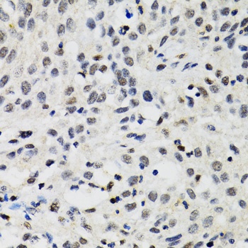
Immunohistochemical staining of human colon cancer tissues using SMARCAD1 antibody.
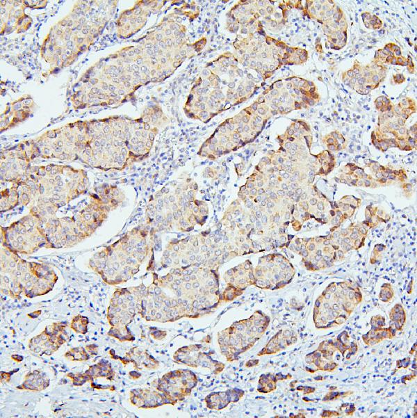
Immunohistochemical staining of human mammary cancer tissues using SMARCAD1 antibody.
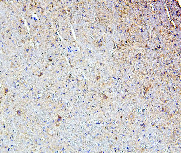
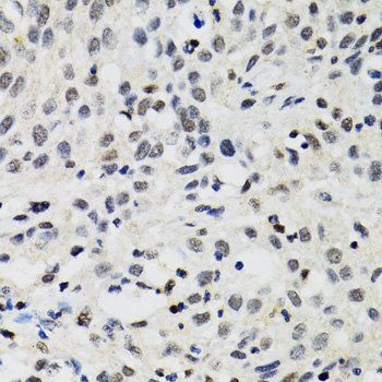
Immunohistochemical staining of mouse brain tissues using SMARCAD1 antibody.
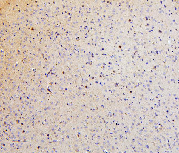
Immunohistochemical staining of rat brain tissues using SMARCAD1 antibody.
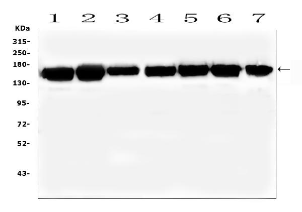
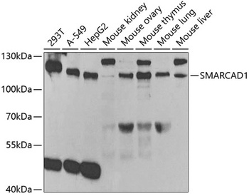
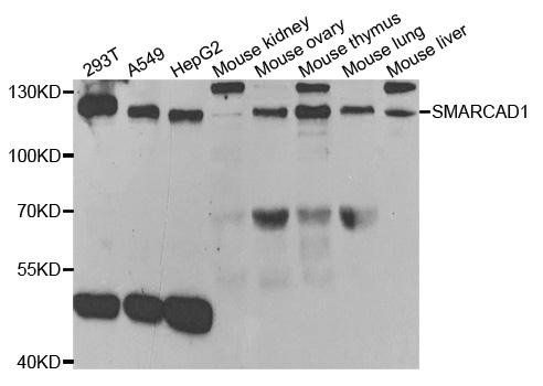
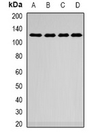
Western blot analysis of Lane 1: human K562 whole cell lysates, Lane 2: human HL-60 whole cell lysates, Lane 3: human U2OS whole cell lysates, Lane 4: human U-87MG whole cell lysates, Lane 5: human HepG2 whole cell lysates, Lane 6: human THP-1 whole cell
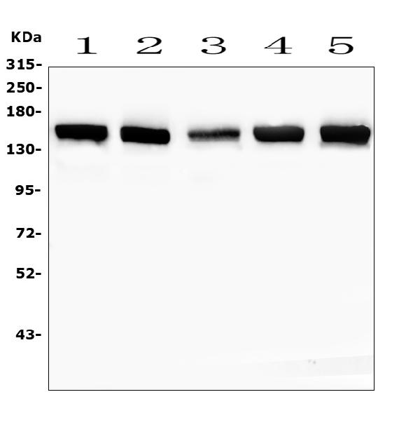
Western blot analysis of Lane 1: rat testicular tissue lysates, Lane 2: mouse thymus tissue lysates, Lane 3: mouse lung tissue lysates, Lane 4: mouse testicular tissue lysates, Lane 5: mouse kidney tissue lysates using SMARCAD1 antibody.
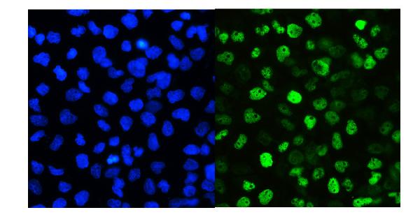
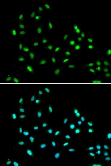
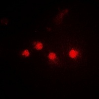
IF ananlysis of A431 cells using SMARCAD1 antibody.
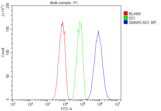
Flow Cytometry analysis of SiHa cells using SMARCAD1 antibody
- Item 1 of 3
- Item 1 of 3
SMARCAD1 antibody [orb178578]
ICC, IF, WB
Human, Mouse, Rat
Polyclonal
Unconjugated
200 μl, 100 μl, 50 μl - Item 1 of 2
SMARCAD1 antibody [orb382045]
IF, WB
Human, Mouse, Rat
Rabbit
Polyclonal
Unconjugated
200 μl, 100 μl, 50 μl - Item 1 of 2
- Item 1 of 2


Submit a review
Filter by Rating

- 5 stars

- 4 stars
- 3 stars
- 2 stars
- 1 stars