You have no items in your shopping cart.
Cart summary
Item 1 of 7
Item 1 of 7
SLC16A11 Antibody
Catalog Number: orb1270501
| Catalog Number | orb1270501 |
|---|---|
| Category | Antibodies |
| Description | SLC16A11 Antibody |
| Species/Host | Rabbit |
| Clonality | Polyclonal |
| Tested applications | FC, IHC-P, WB |
| Reactivity | Human, Mouse |
| Isotype | Rabbit Ig |
| Immunogen | This SLC16A11 antibody is generated from rabbits immunized with a KLH conjugated synthetic peptide between 48-76 amino acids from the N-terminal region of human SLC16A11. |
| Concentration | batch dependent |
| Dilution range | For FACS starting dilution is: 1:25For IHC-P starting dilution is: 1:25For WB starting dilution is: 1:500 |
| Form/Appearance | Liquid |
| Conjugation | Unconjugated |
| MW | 48 kDa |
| Target | SLC16A11 |
| UniProt ID | Q8NCK7 |
| NCBI | Q8NCK7 |
| Storage | Store at 4°C for three months and -20°C, stable for up to one year. As with all antibodies care should be taken to avoid repeated freeze thaw cycles. Antibodies should not be exposed to prolonged high temperatures. |
| Buffer/Preservatives | Supplied in PBS with 0.09% (W/V) sodium azide. |
| Alternative names | Monocarboxylate transporter 11, MCT 11, Solute car Read more... |
| Note | For research use only |
| Application notes | For FACS starting dilution is: 1:25For IHC-P starting dilution is: 1:25For WB starting dilution is: 1:500 |
| Expiration Date | 12 months from date of receipt. |
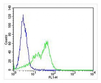
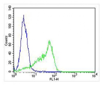
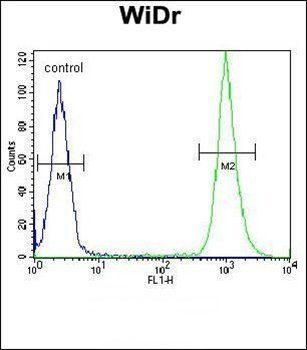
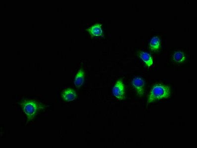
Overlay histogram showing HT-29 cells stained with Antibody (green line). The cells were fixed with 4% paraformaldehyde (10 min) and then permeabilized with 90% methanol for 10 min. The cells were then icubated in 2% bovine serum albumin to block non-specific protein-protein interactions followed by the antibody (1:25 dilution) for 60 min at 37oC. The secondary antibody used was Alexa Fluor 488 goat anti-rabbit lgG (H+L) (1583138) at 1/400 dilution for 40 min at 37oC. Isotype control antibody (blue line) was rabbit IgG1 (1 ug/1x10^6 cells) used under the same conditions. Acquisition of >10000 events was performed.
Overlay histogram showing HT-29 cells stained with Antibody (green line). The cells were fixed with 4% paraformaldehyde (10 min) and then permeabilized with 90% methanol for 10 min. The cells were then icubated in 2% bovine serum albumin to block non-specific protein-protein interactions followed by the antibody (1:25 dilution) for 60 min at 37oC. The secondary antibody used was Alexa Fluor 488 goat anti-rabbit lgG (H+L) (1583138) at 1/400 dilution for 40 min at 37oC. Isotype control antibody (blue line) was rabbit IgG1 (1 ug/1x10^6 cells) used under the same conditions. Acquisition of >10000 events was performed.
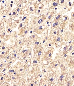
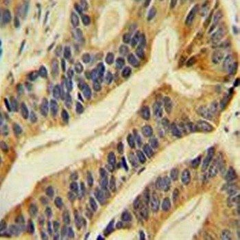
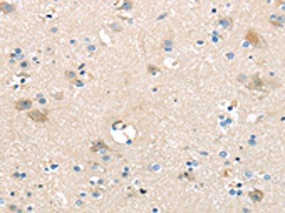
Antibody staining SLC16A11 in Human liver tissue sections by Immunohistochemistry (IHC-P - paraformaldehyde-fixed, paraffin-embedded sections).

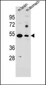
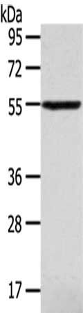
Western Blot at 1:1000 dilution + mouse liver lysates Lysates/proteins at 20 ug per lane.
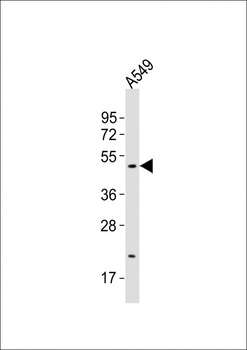
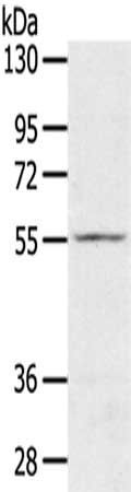
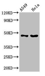
Western Blot at 1:500 dilution + A549 whole cell lysates Lysates/proteins at 20 ug per lane.

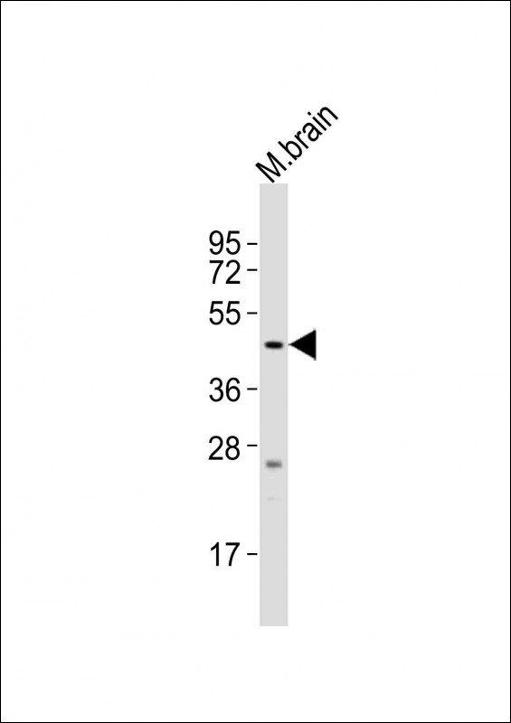
Western Blot at 1:2000 dilution + mouse brain lysates Lysates/proteins at 20 ug per lane.
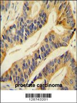
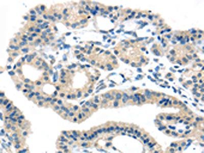
SLC16A11 Antibody immunohistochemistry analysis in formalin fixed and paraffin embedded human prostate carcinoma followed by peroxidase conjugation of the secondary antibody and DAB staining.
- Item 1 of 6
- Item 1 of 3
- Item 1 of 2
- Item 1 of 1


Submit a review
Filter by Rating

- 5 stars

- 4 stars
- 3 stars
- 2 stars
- 1 stars