You have no items in your shopping cart.
Cart summary
SHANK3 antibody
Catalog Number: orb148741
| Catalog Number | orb148741 |
|---|---|
| Category | Antibodies |
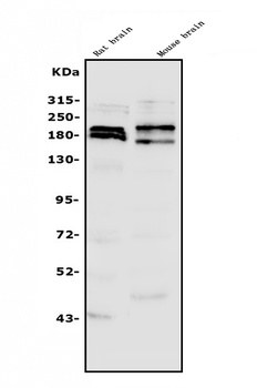
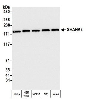
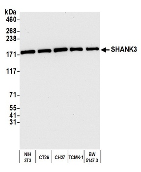
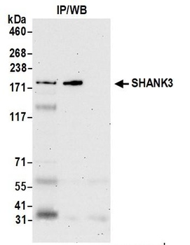
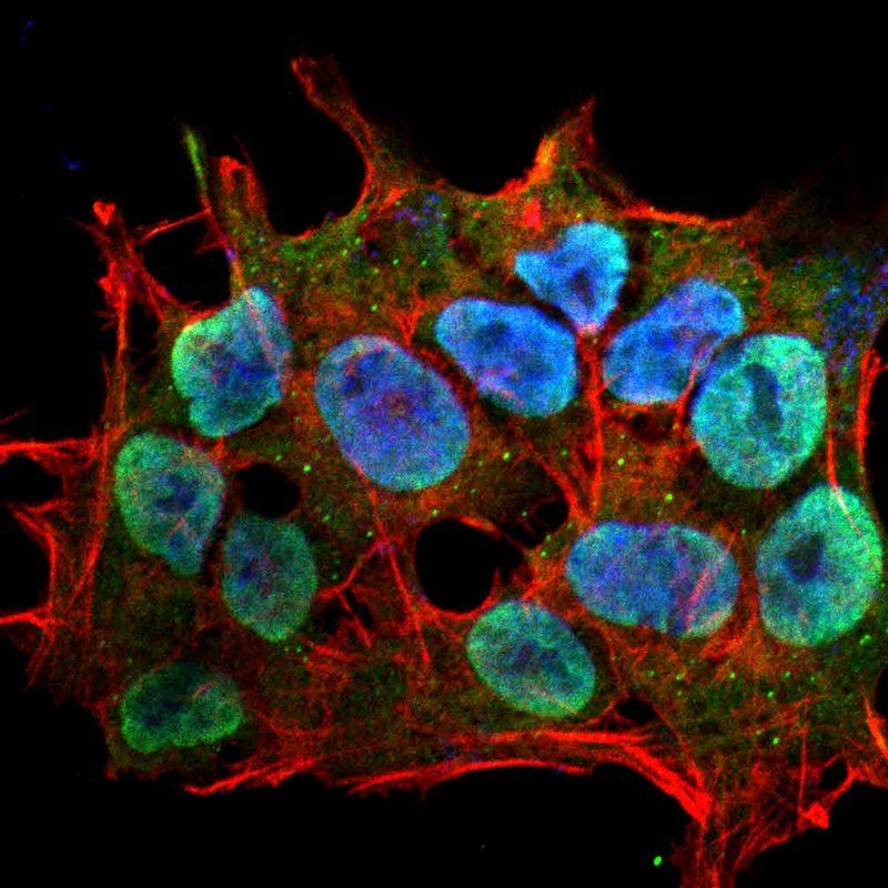
| Description | Mouse monoclonal to SHANK3 (HRP). Shank proteins make up a family of scaffold proteins identified through their interaction with a variety of membrane and cytoplasmic proteins. Shank proteins at postsynaptic sites of excitatory synapses play roles in signal transmission into the postsynaptic neuron. Shank proteins are also crucial in receptor tyrosine kinase signaling; specifically, Shank3 can mediate Erk-MAPK and P13K signaling which is crucial for tubule formation. Shank3 is also one of the latest genes to be associated with autism. A mutation of a single copy of Shank3 on chromosome 22q13 can result in language and/or social communication disorders.. |
| Species/Host | Mouse |
| Clonality | Monoclonal |
| Clone Number | S69 |
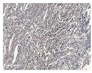
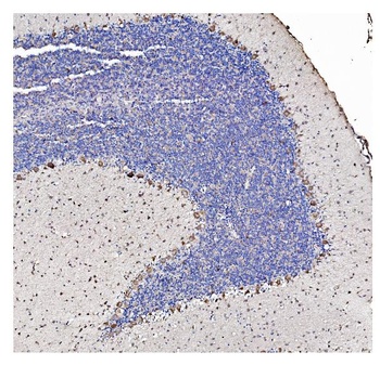
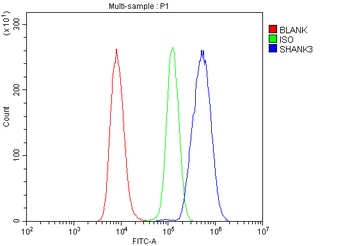
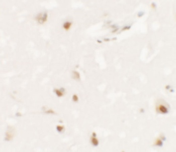
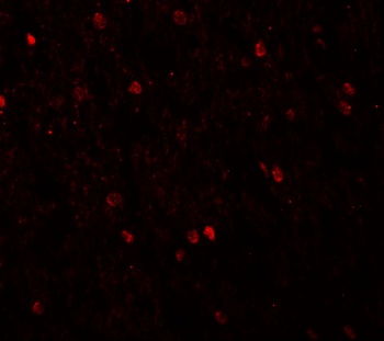
| Tested applications | AM, ICC, IHC, IP, WB |
| Reactivity | Human, Mouse, Rat |
| Isotype | IgG2b |
| Immunogen | Synthetic peptide amino acids 840-857 of rat Shank3 |
| Concentration | 1 mg/ml |
| Dilution range | WB (1:1000), IHC (1:100), ICC/IF (1:100) |
| Conjugation | HRP |
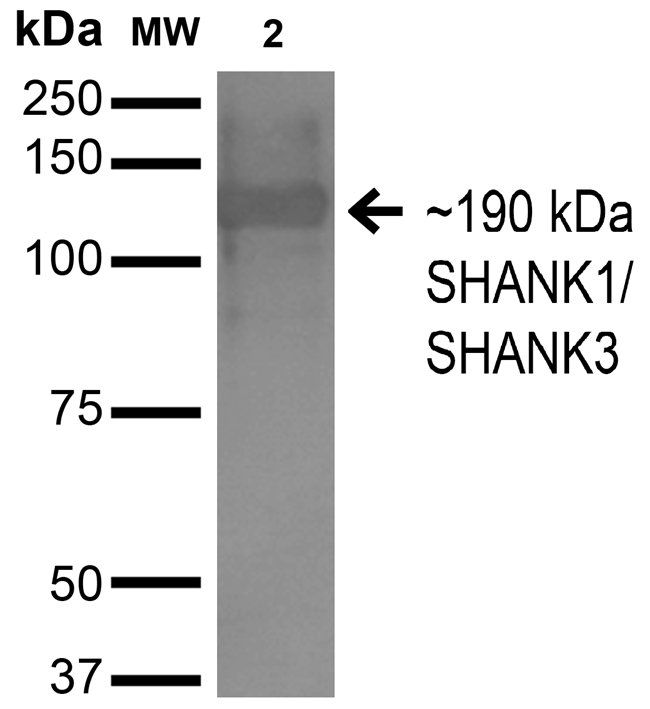
| MW | 190kDa |
| Target | SHANK3 |
| Entrez | 59312 |
| UniProt ID | Q9JLU4 |
| NCBI | NP_067708.1 |
| Storage | Conjugated antibodies should be stored at 4°C |
| Buffer/Preservatives | PBS pH 7.4, 50% glycerol, 0.09% sodium azide *Storage buffer may change when conjugated |
| Alternative names | AI841104 antibody, DEL22q13.3 antibody, KIAA1650 a Read more... |
| Note | For research use only |
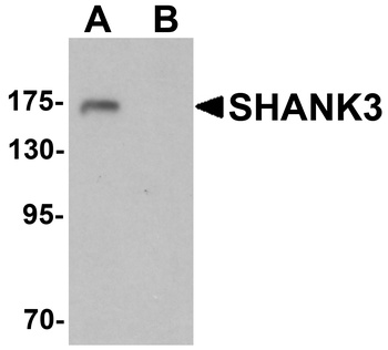
| Application notes | 1 µg/ml of SMC-336 was sufficient for detection of Shank3 in 10 µg COS cell lysate transiently transfected with Shank3 by colorimetric immunoblot analysis using goat anti-mouse IgG:HRP as the secondary antibody. |
| Expiration Date | 12 months from date of receipt. |
- Item 1 of 6
SHANK3 Antibody [orb669123]
ELISA, FC, IHC, WB
Human, Mouse, Rat
Rabbit
Polyclonal
Unconjugated
10 μg, 100 μg - Item 1 of 3
SHANK3 Antibody [orb1238987]
ELISA, IF, IHC-P, WB
Human, Mouse
Rabbit
Polyclonal
Unconjugated
0.1 mg, 0.02 mg - Item 1 of 3
- Item 1 of 3
- Item 1 of 2


Submit a review
Filter by Rating

- 5 stars

- 4 stars
- 3 stars
- 2 stars
- 1 stars