You have no items in your shopping cart.
Cart summary
Item 1 of 7
Item 1 of 7
RYK Antibody
Catalog Number: orb1263009

| Catalog Number | orb1263009 |
|---|---|
| Category | Antibodies |
| Description | RYK Antibody |
| Target | RYK |
| Clonality | Polyclonal |
| Isotype | Rabbit Ig |
| Conjugation | Unconjugated |
| Reactivity | Human, Mouse, Rat |
| Form/Appearance | Liquid |
| Concentration | batch dependent |
| Buffer/Preservatives | Supplied in PBS with 0.09% (W/V) sodium azide. |
| Immunogen | This RYK antibody is generated from rabbits immunized with a KLH conjugated synthetic peptide between 160-190 amino acids from human RYK. |
| UniProt ID | P34925 |
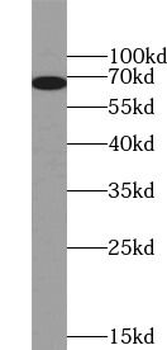
| MW | 68 kDa |
| Tested applications | IHC-P, WB |
| Application notes | For WB starting dilution is: 1:1000For IHC-P starting dilution is: 1:50~100 |
| Antibody Type | Primary Antibody |
| Storage | Maintain refrigerated at 2-8°C for up to 2 weeks. For long term storage store at -20°C in small aliquots to prevent freeze-thaw cycles. |
| Alternative names | Tyrosine-protein kinase RYK, RYK, JTK5A |
| Note | For research use only |
| NCBI | P34925 |
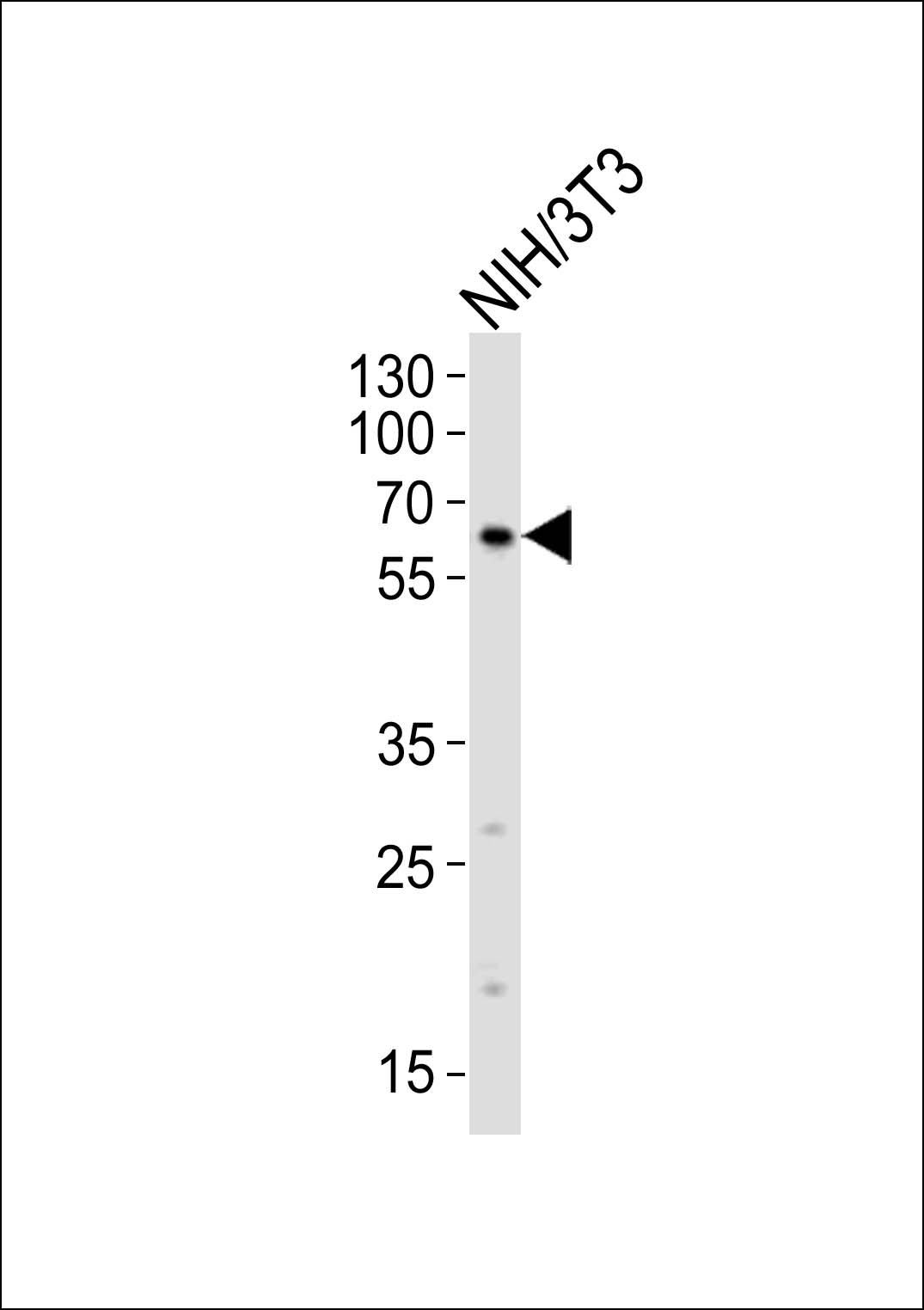
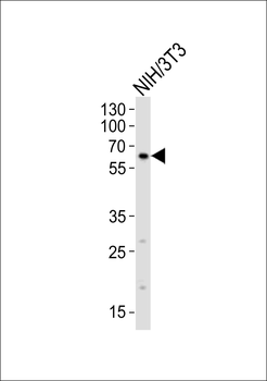
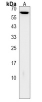
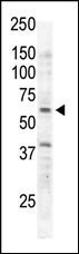
Western blot analysis of lysate from mouse NIH/3T3 cell line, using RYK Antibody (N175) at 1:1000.

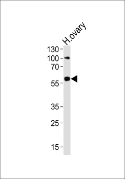
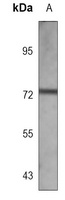
Western blot analysis of lysate from human ovary tissue lysate, using RYK Antibody (N175) at 1:1000.
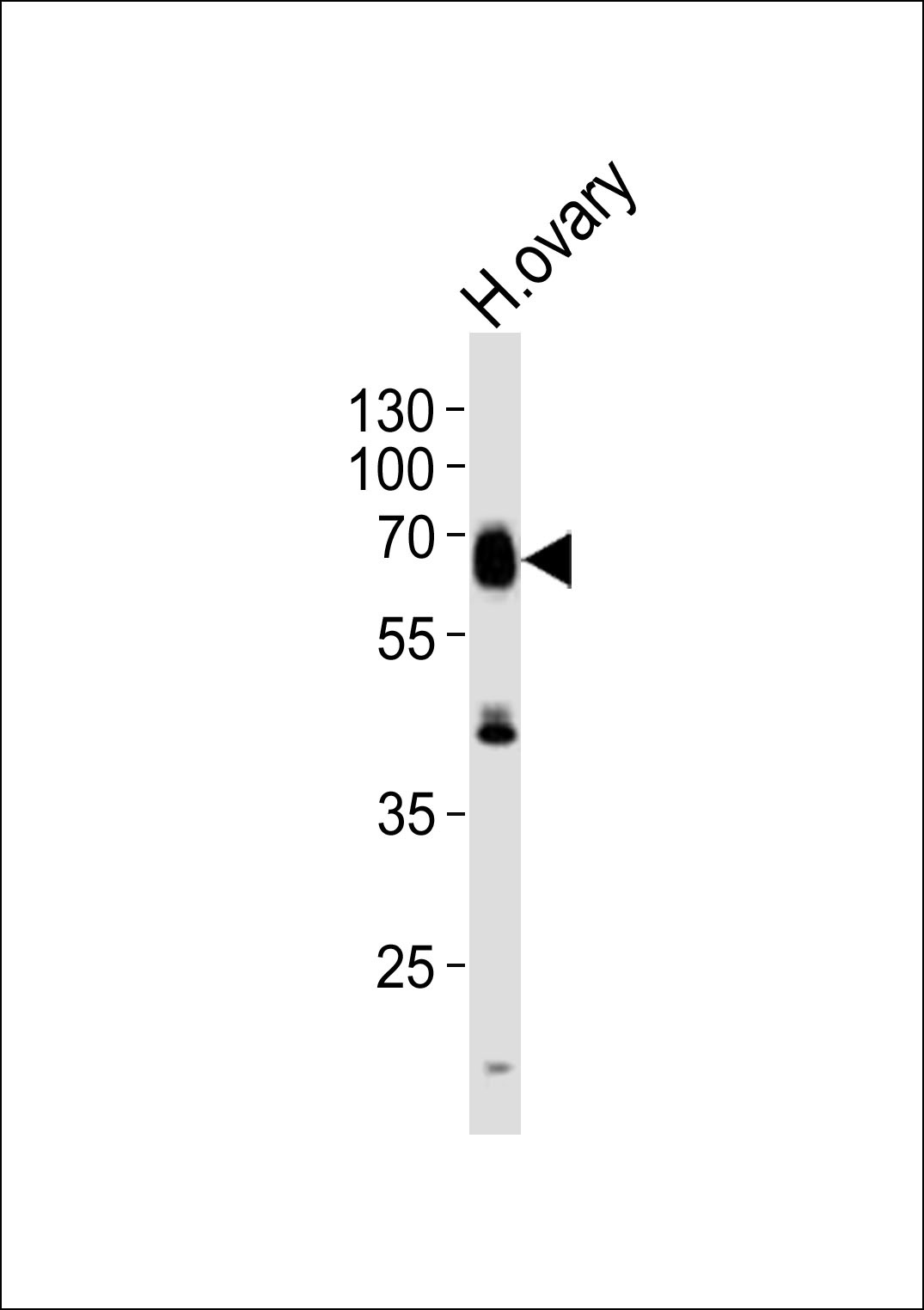
Western blot analysis of lysate from human ovary tissue lysate, using RYK Antibody (N175) at 1:1000.
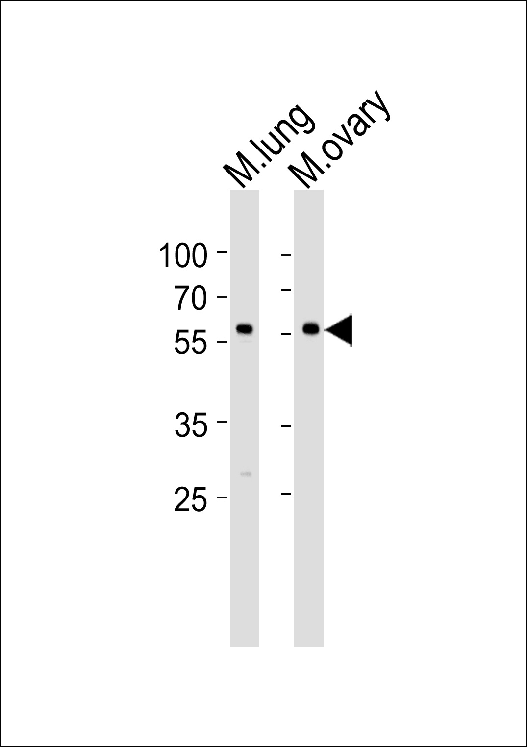
Western blot analysis of lysates from mouse lung and ovary tissue lysate (from left to right), using RYK Antibody (N175) at 1:1000 at each lane.
Antibody is used in Western blot to detect RYK in Jurkat cell lysate.

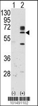
Western blot analysis of RYK using rabbit polyclonal RYK Antibody.293 cell lysates (2 ug/lane) either nontransfected (Lane 1) or transiently transfected with the RYK gene (Lane 2).
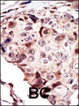
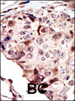
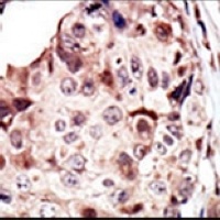
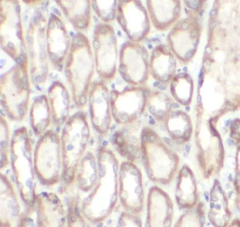
Formalin-fixed and paraffin-embedded human cancer tissue reacted with the primary antibody, which was peroxidase-conjugated to the secondary antibody, followed by AEC staining. BC = breast carcinoma; HC = hepatocarcinoma.
- Item 1 of 7
- Item 1 of 2
- Item 1 of 2
- Item 1 of 3
RYK Antibody [orb630913]
ELISA, IF, IHC, WB
Human, Mouse, Rat
Rabbit
Polyclonal
Unconjugated
100 μg, 50 μg 
Goat anti-Ryk (mouse) Antibody [orb12336]
ELISA
Bovine, Canine, Human, Mouse, Porcine, Rat
Goat
Polyclonal
Unconjugated
100 μg