You have no items in your shopping cart.
Cart summary
Item 1 of 15
Item 1 of 15
Ripk3 Antibody
Catalog Number: orb1239942
| Catalog Number | orb1239942 |
|---|---|
| Category | Antibodies |
| Description | Ripk3 Antibody |
| Species/Host | Rabbit |
| Clonality | Polyclonal |
| Tested applications | ELISA, IF, IHC-P, IP, WB |
| Reactivity | Human, Mouse, Rat |
| Isotype | IgG |
| Immunogen | Anti-RIP3 antibody (orb1239942) was raised against a peptide corresponding to 14 amino acids near the carboxy terminus of murine RIP3. The immunogen is located within the last 50 amino acids of RIP3. |
| Concentration | 1 mg/ml |
| Dilution range | WB: 0.1-0.5 μg/mL; IHC-P: 5 μg/mL; IF: 20 μg/mL, IP: 20 μg/mL.Antibody validated: Western Blot in mouse and human samples; Immunohistochemistry in mouse and rat samples; Immunofluorescence in mouse and rat samples; Immunoprecipitation in human samples; Immunogold EM in mouse samples. All other applications and species not yet tested. |
| Form/Appearance | Liquid |
| Conjugation | Unconjugated |
| MW | Predicted: 53kD for mouse RIP3 and 57kD for human RIP3Observed: 53kD for mouse RIP3 and 57kD for human RIP3 |
| Target | Ripk3 |
| UniProt ID | Q9QZL0 |
| NCBI | AAF03133 |
| Storage | RIP3 antibody can be stored at 4°C up to one year. Antibodies should not be exposed to prolonged high temperatures. |
| Buffer/Preservatives | RIP3 Antibody is supplied in PBS containing 0.02% sodium azide. |
| Alternative names | RIP3 Antibody: Rip3, AW107945, 2610528K09Rik, Rip3 Read more... |
| Note | For research use only |
| Expiration Date | 12 months from date of receipt. |
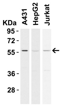
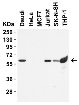
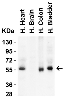
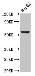
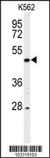
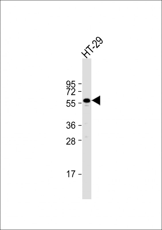
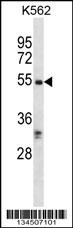
Western Blot Validation in Human Cell Lines. Loading: 15 µg of lysates per lane. Antibodies: RIP3 orb1239942, (0.5 µg/mL), 1h incubation at RT in 5% NFDM/TBST. Secondary: Goat anti-rabbit IgG HRP conjugate at 1:10000 dilution.
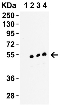
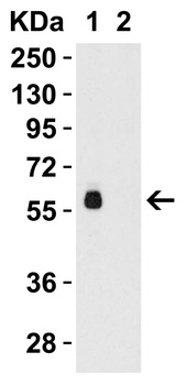
Western Blot Validation in C2C12 Cells Mouse. Loading: 15 µg of lysates per lane. Antibodies: RIP3 orb1239942, 1h incubation at RT in 5% NFDM/TBST. Secondary: Goat anti-rabbit IgG HRP conjugate at 1:10000 dilution. Lane 1: orb1239942, 0.1 µg/mL in the presence of peptide blocking, Lane 2: orb1239942, 0.1 µg/mL, Lane 3: orb1239942, 0.2 µg/mL, Lane 4: orb1239942, 0.5 µg/mL.
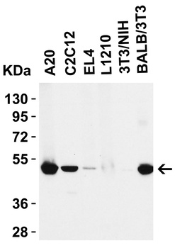
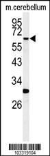
Western Blot Validation in Mouse Cell lines. Loading: 15 µg of lysates per lane. Antibodies: RIP3 orb1239942, (0.5 µg/mL), 1h incubation at RT in 5% NFDM/TBST. Secondary: Goat anti-rabbit IgG HRP conjugate at 1:10000 dilution.
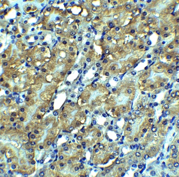
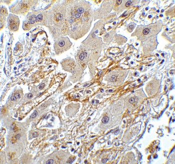
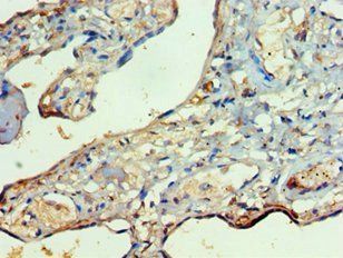
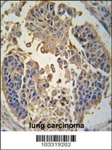
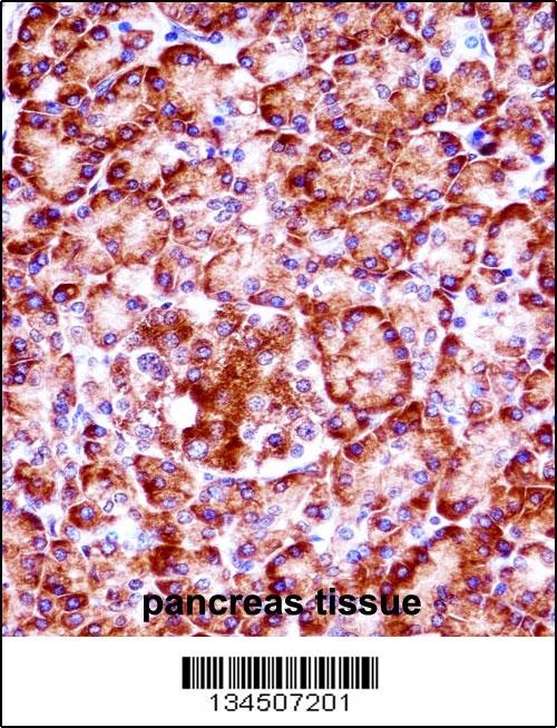
Immunohistochemistry Validation of RIP3 in Mouse Kidney Tissue. Immunohistochemical analysis of paraffin-embedded mouse kidney tissue using anti-RIP3 antibody (orb1239942) at 2.5 µg/ml. Tissue was fixed with formaldehyde and blocked with 10% serum for 1 h at RT; antigen retrieval was by heat mediation with a citrate buffer (pH6). Samples were incubated with primary antibody overnight at 4°C. A goat anti-rabbit IgG H&L (HRP) at 1/250 was used as secondary. Counter stained with Hematoxylin.
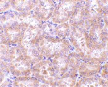
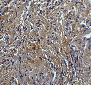
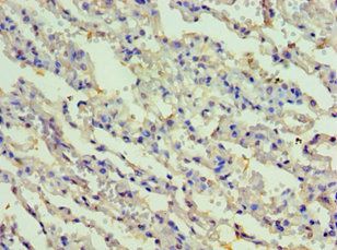
Immunohistochemistry Validation of RIP3 in Rat Kidney Tissue. Immunohistochemical analysis of paraffin-embedded rat kidney tissue using anti-RIP3 antibody (orb1239942) at 5 µg/ml. Tissue was fixed with formaldehyde and blocked with 10% serum for 1 h at RT; antigen retrieval was by heat mediation with a citrate buffer (pH6). Samples were incubated with primary antibody overnight at 4°C. A goat anti-rabbit IgG H&L (HRP) at 1/250 was used as secondary. Counter stained with Hematoxylin.
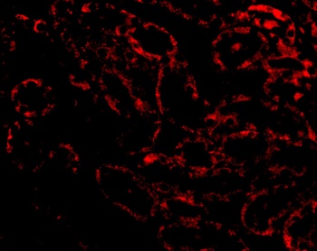
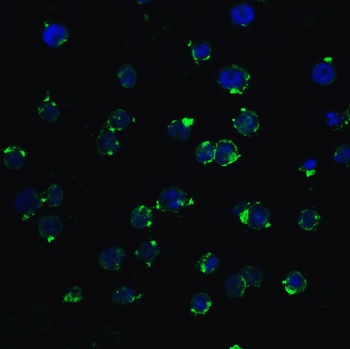
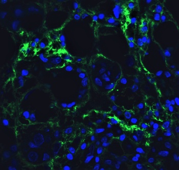
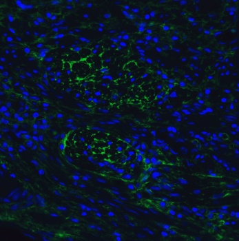
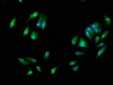
Immunofluorescence Validation of RIP3 in Rat Kidney Tissue. Immunofluorescent analysis of 4% paraformaldehyde-fixed Rat Kidney tissue labeling RIP3 with orb1239942 at 20 µg/mL, followed by goat anti-rabbit IgG secondary antibody at 1/500 dilution (red).
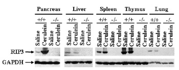
KO Validation of RIP3 in RIP3 KO Mice (He et al., 2009). Western blot analysis of RIP3 with anti-RIP3 antibodies shows disrupted RIP3 expression in pancreas, liver spleen thymus and lung of RIP3 KO mice. Cerulein treatment upregulated RIP3 expression in the pancreas of WT mice.
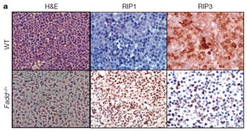
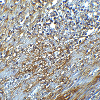
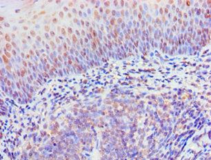
Immunohistochemistry Validation of RIP3 in Fadd KO mice (Zhang et al., 2011). RIP3 expression detected by anti-RIP3 antibodies (orb1239942) was decreased in Fadd KO mice as compared to WT mice.
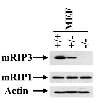
KO Validation of RIP3 in MEF Cells (He et al., 2012). Western blot analysis of RIP3 with anti-RIP3 antibodies shows disrupted RIP3 expression in RIP3 KO MEFs, but not in WT MEF cells.
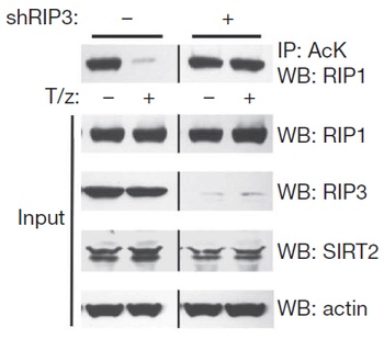
KD Validation of RIP3 in L929 Cells (Narayan et al., 2012). Immunoblot analysis of RIP3 with anti-RIP3 antibodies (orb1239942) shows RIP3 expression was disrupted in RIP3 knockdown L929 cells.
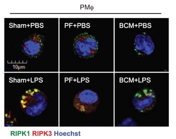
Regulated Expression Validation of RIP3 in Macrophages (Li et al., 2016). Confocal microscopy shows colocalization of RIPK1 (green) and RIPK3 (red) in PMϕ and RIPK3 was detected by anti-RIPK3 antibodies (orb1239942). PF or BCM suppressed LPS-induced upregulation of RIP3.
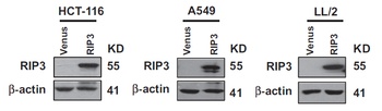
Overexpression Validation of RIP3 in Cancer Cell Lines (Yang et al., 2017). The cancer cell lines were stably expressing flag-tagged RIP3 and RIP expression was detected by anti-RIP3 antibodies (orb1239942) in RIP3-overexpressed cells.
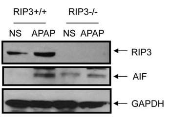
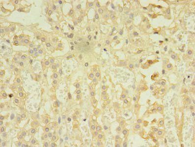
Induced Expression of RIP3 by Acetaminophen in WT and RIP3 KO Mice (Ramachandran et al., 2013). Wild-type and RIP3 KO mice were treated with300 mg/kg acetaminophen or saline. RIP3 expression detected by anti-RIP3 antibodies (orb1239942) were up-regulated after acetaminophen treatment in the liver, while this effect was not observed in RIP3 KO mice.
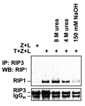
IP Validation of RIP3 in HT29 Cells (Li et al., 2012). RIP3 complexes isolated by immunoprecipitation with anti-RIP3 antibodies (orb1239942) from HT-29 cells treated with T+Z+L after lysis in regular lysis buffer or buffer containing the indicated amount of urea or NaOH.
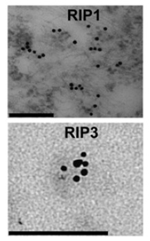
Immunogold EM Validation in MEF Cells (Li et al., 2012). Clustering of RIP3 in necrotic MEFs shown by immunogold EM with anti-RIP3 antibodies (orb1239942). Scale bars, 100 nm (RIP3).
- Item 1 of 9
- Item 1 of 3
- Item 1 of 3
- Item 1 of 3
- Item 1 of 3
Submit a review
Filter by Rating

- 5 stars

- 4 stars
- 3 stars
- 2 stars
- 1 stars