You have no items in your shopping cart.
Cart summary
Item 1 of 5
Item 1 of 5
PSMB1 Antibody
Catalog Number: orb1262482

| Catalog Number | orb1262482 |
|---|---|
| Category | Antibodies |
| Description | PSMB1 Antibody |
| Target | PSMB1 |
| Clonality | Polyclonal |
| Isotype | Rabbit Ig |
| Conjugation | Unconjugated |
| Reactivity | Human, Mouse |
| Form/Appearance | Liquid |
| Concentration | batch dependent |
| Buffer/Preservatives | Supplied in PBS with 0.09% (W/V) sodium azide. |
| Immunogen | This PSMB1 antibody is generated from rabbits immunized with a KLH conjugated synthetic peptide between 214-241 amino acids from the C-terminal region of human PSMB1. |
| UniProt ID | P20618 |
| MW | 26 kDa |
| Tested applications | FC, IF, IHC-P, WB |
| Application notes | For WB starting dilution is: 1:1000For IHC-P starting dilution is: 1:10~50For IF starting dilution is: 1:10~50For FACS starting dilution is: 1:10~50 |
| Antibody Type | Primary Antibody |
| Storage | Maintain refrigerated at 2-8°C for up to 2 weeks. For long term storage store at -20°C in small aliquots to prevent freeze-thaw cycles. |
| Alternative names | Proteasome subunit beta type-1, Macropain subunit Read more... |
| Note | For research use only |
| NCBI | P20618 |

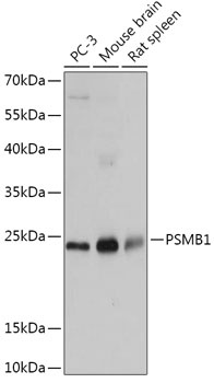
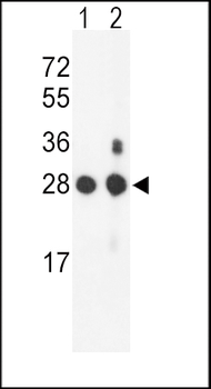
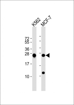
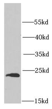
Western Blot at 1:1000 dilution Lane 1: K562 whole cell lysate Lane 2: MCF-7 whole cell lysate Lysates/proteins at 20 ug per lane.

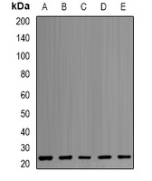
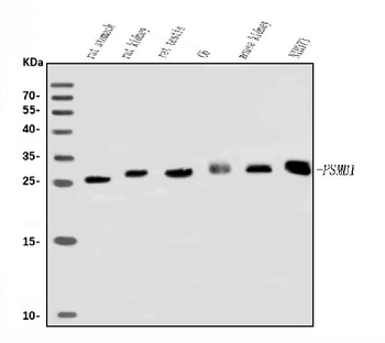
Western blot analysis of PSMB1 Antibody in mouse NIH-3T3 cell line (lane 1) and mouse bladder tissue (lane 2) lysates (35 ug/lane)

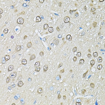
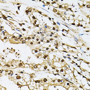
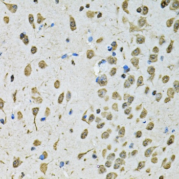
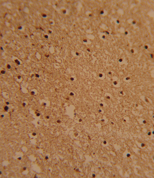
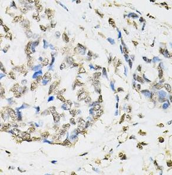
Formalin-fixed and paraffin-embedded human brain tissue reacted with PSMB1 Antibody, which was peroxidase-conjugated to the secondary antibody, followed by DAB staining.
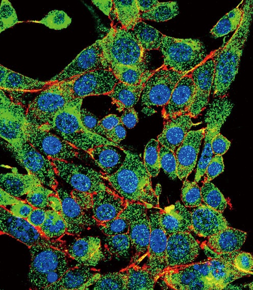
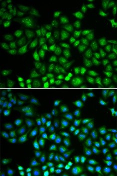
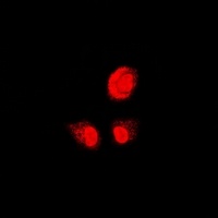
Confocal immunofluorescent analysis of PSMB1 Antibody with HepG2 cell followed by Alexa Fluor 488-conjugated goat anti-rabbit lgG (green). Actin filaments have been labeled with Alexa Fluor 555 phalloidin (red). DAPI was used to stain the cell nuclear (blue).
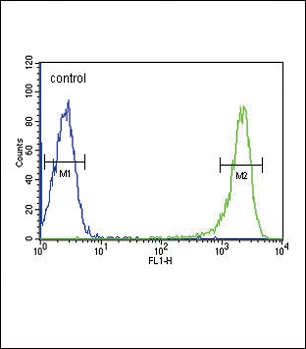
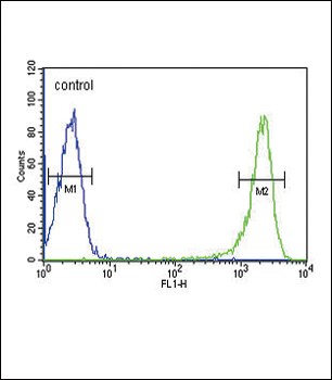
Flow cytometric analysis of HL-60 cells (right histogram) compared to a negative control cell (left histogram). FITC-conjugated goat-anti-rabbit secondary antibodies were used for the analysis.
- Item 1 of 5
- Item 1 of 5
PSMB1 Antibody (C-term) [orb1928242]
FC, IF, IHC-P, WB
Human, Mouse
Rabbit
Polyclonal
Unconjugated
100 μl, 50 μl - Item 1 of 2
Anti-PSMB1 Antibody [orb340722]
IF, WB
Human, Mouse, Rat
Rabbit
Polyclonal
Unconjugated
200 μl, 100 μl, 50 μl - Item 1 of 2
Anti-Psmb1 Antibody [orb763073]
ELISA, FC, WB
Human, Mouse, Rat
Rabbit
Polyclonal
Unconjugated
100 μg, 10 μg - Item 1 of 2
PSMB1 Antibody [orb630294]
ELISA, IF, IHC, WB
Human, Mouse, Rat
Rabbit
Polyclonal
Unconjugated
100 μg, 50 μg