You have no items in your shopping cart.
Protein A Alkaline Phosphatase Conjugated
SKU: orb348751
Description
Research Area
Signal Transduction
Images & Validation
−Item 1 of 2
| Application Notes |
|---|
Key Properties
−| Purity | Protein A Alkaline Phosphatase Conjugated was prepared from chromatographically pure Protein A. Protein A Alkaline Phosphatase Conjugated assayed by immunoelectrophoresis resulted in a single precipitin arc against anti-Alkaline Phosphatase (calf intestine) and anti-Protein A. No reaction was observed against anti-Protein G. |
|---|---|
| Conjugation | AP |
Storage & Handling
−| Storage | Store vial at 4° C before opening. DO NOT FREEZE. Protein A Alkaline Phosphatase Conjugated is stable at 4° C as an undiluted liquid. Dilute only prior to immediate use. Freezing alkaline phosphatase conjugates will result in a substantial loss of enzymatic activity. |
|---|---|
| Form/Appearance | Liquid (sterile filtered) |
| Buffer/Preservatives | Preservative: 0.1% (w/v) Sodium Azide. Stabilizer: 10 mg/mL Bovine Serum Albumin (rAlbumin) - Immunoglobulin and Protease free; Buffer: 0.05 M Tris Chloride, 0.15M Sodium Chloride, 0.001M Magnesium Chloride, 0.0001M Zinc Chloride, 50% (v/v) Glycerol; pH 8.0 |
| Concentration | 1.0 mg/mL |
| Expiration Date | 6 months from date of receipt. |
| Hazard Information | Non-Toxic |
| Disclaimer | For research use only |
Alternative Names
−ProA, Staphylococcus A protein, ALP, Protein A Alk Phos
Similar Products
−- Item 1 of 1
ALPP Mouse Monoclonal Antibody [orb1474075]
WB
Human
Mouse
Monoclonal
Unconjugated
100 μl, 50 μl, 200 μl, 30 μl 
Quality Guarantee
Explore bioreagents carefree to elevate your research. All our products are rigorously tested for performance. If a product does not perform as described on its datasheet, our scientific support team will provide expert troubleshooting, a prompt replacement, or a refund. For full details, please see our Terms & Conditions and Buying Guide. Contact us at [email protected].
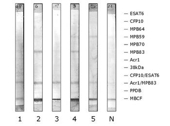
Antibody responses to multiple Mycobacterium tuberculosis-complex antigens in a meerkat with Mycobacterium bovis infection revealed by multiantigen print immunoassay (MAPIA) of sequential serum samples. Incubated with alkaline phosphatase-conjugated protein A (p/n orb348751). Antigens are indicated in the right margin, with a band visible on each strip where antibody is present. Samples were collected at the following time points: 1 = 0 months (December 2005); 2 = 8 months (August 2006); 3 = 10 months (October 2006); 4 = 15 months (March 2007); 5 = 17 months (May 2007). N = negative control.

Rapid test (lateral flow immunoassay) results of sequential serum samples from a meerkat with Mycobacterium bovis infection. In the test window, the upper line (labeled C) is an internal positive control indicating mobility of the serum sample and diluent across the membrane. The lower bands of reactivity (labeled T) visualized on samples 1–5 indicate a positive reaction to the test antigens. Relative test band strengths are given in brackets: 1 = 0 months (+); 2 = 8 months (++); 3 = 10 months (+++); 4 = 15 months (++); 5 = 17 months (+++). N = negative control. Tracheal washes collected from the same animal at time points 1 and 5 cultured positive for M. bovis.
Quick Database Links
UniProt
UniProt Details
− No UniProt data available
Documents Download
Datasheet
Product Information
Request a Document
Protocol Information
WB
Western Blot (IB, immunoblot)
IHC
Immunohistochemistry
ELISA
Enzyme-linked Immunosorbent Assay (EIA)
DOT
Dot Blot
Protein A Alkaline Phosphatase Conjugated (orb348751)

- 0.0
Based on 0 reviews
Participating in our Biorbyt product reviews program enables you to support fellow scientists by sharing your firsthand experience with our products.
Login to Submit a ReviewAvailable Sizes
Select a size below