You have no items in your shopping cart.
Cart summary
Item 1 of 5
Item 1 of 5
PMEL17 / Melanoma gp100 Antibody
Catalog Number: orb606653

| Catalog Number | orb606653 |
|---|---|
| Category | Antibodies |
| Description | Cytotoxic T lymphocytes (CTL's) recognize melanoma-associated antigens, which belong to three main groups. These groups include tumor-associated testis-specific antigens, melanocyte differentiation antigens and mutated or aberrantly expressed antigens, which are routinely used as markers to identify melanomas based on their binding to specific monoclonal antibodies. gp100, also designated ME20-M, ME20-S and PMEL 17, is classified as a melanocyte differentiation antigen and is expressed at low levels in normal cell lines and tissues, but is upregulated in melanocytes. gp100 is a highly glycosylated protein. It is also the product of proteolytic cleavage, which results in a secreted protein.The gp100 molecule is a 100kDa glycosylated protein that is cleaved into a small (26kDa) carboxy-terminal fragment and a larger amino- terminal section (60-64 kDa), which is subsequently cleaved to generate 26kDa and 34-38kDa fragments. |
| Clonality | Monoclonal |
| Species/Host | Mouse |
| Isotype | Mouse IgG1, kappa |
| Conjugation | Unconjugated |
| Reactivity | Human |
| Buffer/Preservatives | 0.2 mg/ml in 1X PBS with 0.1 mg/ml rAlbumin (US sourced) and 0.05% sodium azide |
| Purity | Protein G affinity chromatography |
| Immunogen | A portion of amino acids 376-502 from the human protein was used as the immunogen for the PMEL17 antibody. |
| UniProt ID | P40967 |
| Tested applications | ELISA, IHC-P, WB |
| Dilution range | ELISA: 2-4ug/ml (order BSA/azide-free format),Immunohistochemistry (FFPE): 1-2ug/ml for 30 min at RT,Western blot: 1-2ug/ml |
| Application notes | Optimal dilution of the PMEL17 antibody should be determined by the researcher. |
| Antibody Type | Primary Antibody |
| Clone Number | PMEL/2037 |
| Formula | 0.2 mg/ml in 1X PBS with 0.1 mg/ml BSA (US sourced) and 0.05% sodium azide |
| Storage | Store the PMEL17 antibody at 2-8°C (with azide) or aliquot and store at -20°C or colder (without azide). |
| Hazard Information | This PMEL17 antibody is available for research use only. |
| Note | For research use only |

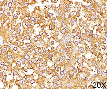
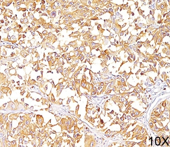
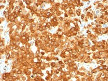
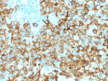
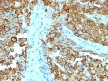
IHC testing of human melanoma with PMEL17 antibody (clone PMEL/2037). Required HIER: boil tissue sections in 10mM citrate buffer, pH6, for 10-20 min followed by cooling at RT for 20 min.
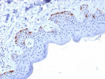
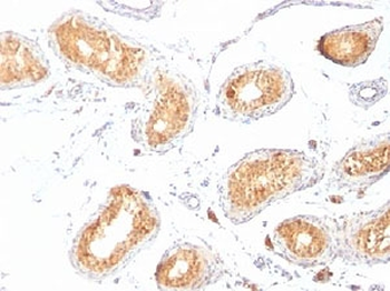
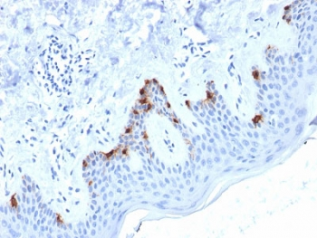
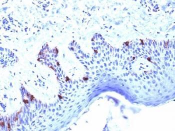
IHC testing of human skin with PMEL17 antibody (clone PMEL/2037). Required HIER: boil tissue sections in 10mM citrate buffer, pH6, for 10-20 min followed by cooling at RT for 20 min.
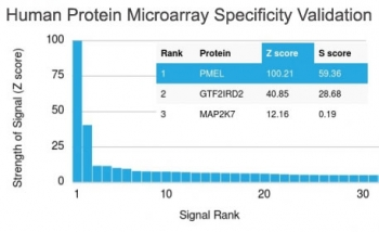
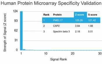
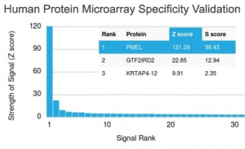
Analysis of HuProt (TM) microarray containing more than 19, 000 full-length human proteins using PMEL17 antibody (clone PMEL/2037). These results demonstrate the foremost specificity of the PMEL/2037 mAb. Z- and S- score: The Z-score represents the strength of a signal that an antibody (in combination with a fluorescently-tagged anti-IgG secondary Ab) produces when binding to a particular protein on the HuProt (TM) array. Z-scores are described in units of standard deviations (SD's) above the mean value of all signals generated on that array. If the targets on the HuProt (TM) are arranged in descending order of the Z-score, the S-score is the difference (also in units of SD's) between the Z-scores. The S-score therefore represents the relative target specificity of an Ab to its intended target.
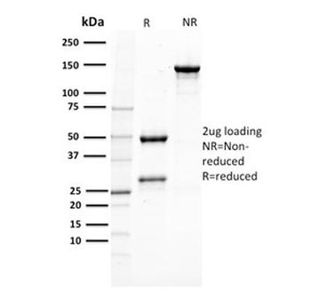
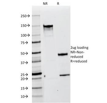
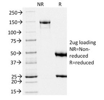
SDS-PAGE analysis of purified, BSA-free PMEL17 antibody (clone PMEL/2037) as confirmation of integrity and purity.
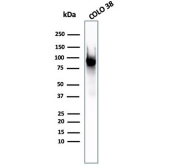
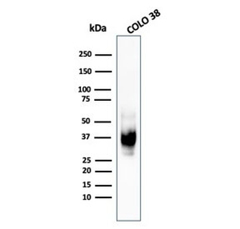
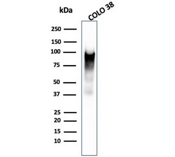
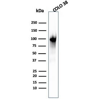
Western blot testing of human COLO-38 cell lysate with PMEL17 antibody (clone PMEL/2037).
- Item 1 of 6
PMEL17 / Melanoma gp100 Antibody [orb248538]
FACS, IF, IHC-P, WB
Human
Mouse
Monoclonal
Unconjugated
20 μg - Item 1 of 6
PMEL17 / Melanoma gp100 Antibody [orb2637927]
FACS, IF, IHC-P, WB
Human
Mouse
Monoclonal
Unconjugated
100 μg - Item 1 of 5
- Item 1 of 5
- Item 1 of 5
PMEL17 / Melanoma gp100 Antibody [orb2641371]
ELISA, IHC-P, WB
Human
Mouse
Monoclonal
Unconjugated
100 μg