You have no items in your shopping cart.
Cart summary
Item 1 of 6
Item 1 of 6
PLA2G7 Antibody (Center)
Catalog Number: orb1927707

| Catalog Number | orb1927707 |
|---|---|
| Category | Antibodies |
| Description | Affinity Purified Rabbit Polyclonal Antibody (Pab) |
| Species/Host | Rabbit |
| Clonality | Polyclonal |
| Clone Number | RB21322 |
| Tested applications | FC, IHC-P, WB |
| Reactivity | Human |
| Isotype | Rabbit IgG |
| Antibody Type | Primary Antibody |
| Dilution range | WB: 1:2000, WB: 1:1000, IHC-P: 1:25, IHC-P: 1:25, IHC-P: 1:10~50, FC: 1:25 |
| Form/Appearance | Purified polyclonal antibody supplied in PBS with 0.09% (W/V) sodium azide. This antibody is purified through a protein A column, followed by peptide affinity purification. |
| Conjugation | Unconjugated |
| MW | 50077 Da |
| Target | This PLA2G7 antibody is generated from rabbits immunized with a KLH conjugated synthetic peptide between 200-228 amino acids from the Central region of human PLA2G7. |
| UniProt ID | Q13093 |
| NCBI | NP_001161829.1, NP_005075.3 |
| Storage | Maintain refrigerated at 2-8°C for up to 2 weeks. For long term storage store at -20°C in small aliquots to prevent freeze-thaw cycles |
| Alternative names | Platelet-activating factor acetylhydrolase, PAF ac Read more... |
| Note | For research use only |
| Expiration Date | 12 months from date of receipt. |
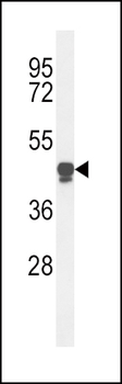
Western blot analysis of PLA2G7 Antibody (Center) in HL-60 cell line lysates (35 ug/lane). PLA2G7 (arrow) was detected using the purified Pab.
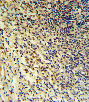
PLA2G7 Antibody (Center) IHC analysis in formalin fixed and paraffin embedded tonsil followed by peroxidase conjugation of the secondary antibody and DAB staining. This data demonstrates the use of the PLA2G7 Antibody (Center) for immunohistochemistry. Clinical relevance has not been evaluated.
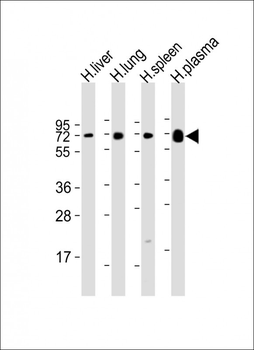
All lanes: Anti-PLA2G7 Antibody (Center) at 1:2000 dilution. Lane 1: human liver lysate. Lane 2: human lung lysate. Lane 3: human spleen lysate. Lane 4: human plasma lysate. Lysates/proteins at 20 µg per lane. Secondary Goat Anti-Rabbit IgG, (H+L), Peroxidase conjugated at 1/10000 dilution. Predicted band size: 50 kDa. Blocking/Dilution buffer: 5% NFDM/TBST.
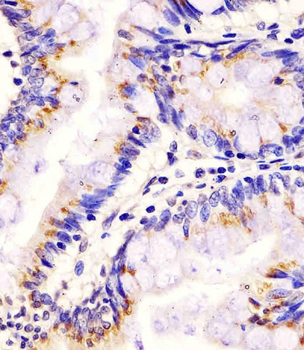
Staining PLA2G7 in human colon tissue sections by Immunohistochemistry (IHC-P - paraformaldehyde-fixed, paraffin-embedded sections). Tissue was fixed with formaldehyde and blocked with 3% BSA for 0.5 hour at room temperature; antigen retrieval was by heat mediation with a citrate buffer (pH6). Samples were incubated with primary antibody (1/25) for 1 hours at 37°C. A undiluted biotinylated goat polyvalent antibody was used as the secondary antibody.
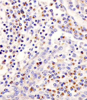
Staining PLA2G7 in human tonsil tissue sections by Immunohistochemistry (IHC-P - paraformaldehyde-fixed, paraffin-embedded sections). Tissue was fixed with formaldehyde and blocked with 3% BSA for 0.5 hour at room temperature; antigen retrieval was by heat mediation with a citrate buffer (pH6). Samples were incubated with primary antibody (1/25) for 1 hours at 37°C. A undiluted biotinylated goat polyvalent antibody was used as the secondary antibody.
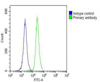
Overlay histogram showing HL-60 cells stained (green line). The cells were fixed with 2% paraformaldehyde (10 min) and then permeabilized with 90% methanol for 10 min. The cells were then icubated in 2% bovine serum albumin to block non-specific protein-protein interactions followed by the antibody (1:25 dilution) for 60 min at 37°C. The secondary antibody used was Goat-Anti-Rabbit IgG, DyLight 488 Conjugated Highly Cross-Adsorbed at 1/200 dilution for 40 min at 37°C. Isotype control antibody (blue line) was rabbit IgG (1 μg/1x10^6 cells) used under the same conditions. Acquisition of > 10000 events was performed.
