You have no items in your shopping cart.
Cart summary
Item 1 of 5
Item 1 of 5
PKC iota Antibody / PRKCI
Catalog Number: orb2636969

| Catalog Number | orb2636969 |
|---|---|
| Category | Antibodies |
| Description | Members of the protein kinase C (PKC) family play a key regulatory role in a variety of cellular functions, including cell growth and differentiation, gene expression, hormone secretion and membrane function. PKCs were originally identified as serine/threonine protein kinases whose activity was dependent on calcium and phospholipids. Diacylglycerols (DAG) and tumor promoting phorbol esters bind to and activate PKC. PKCs can be subdivided into at least two major classes, including conventional (c) PKC isoforms (, , , l/i, m and n). Patterns of expression for each PKC isoform differ among tissues and PKC family members exhibit clear differences in their cofactor dependencies. For instance, the kinase activities of PKC �L and are independent of Ca2+. On the other hand, most of the other PKC members possess phorbol ester-binding activities and kinase activities. |
| Species/Host | Mouse |
| Clonality | Monoclonal |
| Clone Number | PRKCI/4911 |
| Tested applications | IHC-P |
| Reactivity | Human |
| Isotype | Mouse IgG2b, kappa |
| Immunogen | A portion of amino acids 100-300 was used as the immunogen for the PRKCI antibody. |
| Antibody Type | Primary Antibody |
| Dilution range | Immunohistochemistry (FFPE): 1-2ug/ml |
| Purity | Protein A/G affinity |
| Conjugation | Unconjugated |
| Formula | 0.2 mg/ml in 1X PBS with 0.1 mg/ml BSA (US sourced), 0.05% sodium azide |
| Hazard Information | This PRKCI antibody is available for research use only. |
| UniProt ID | P41743 |
| Storage | Maintain refrigerated at 2-8°C for up to 2 weeks. For long term storage store at -20°C in small aliquots to prevent freeze-thaw cycles. |
| Buffer/Preservatives | 0.2 mg/ml in 1X PBS with 0.1 mg/ml rAlbumin (US sourced), 0.05% sodium azide |
| Note | For research use only |
| Application notes | Optimal dilution of the PRKCI antibody should be determined by the researcher. |
| Expiration Date | 12 months from date of receipt. |

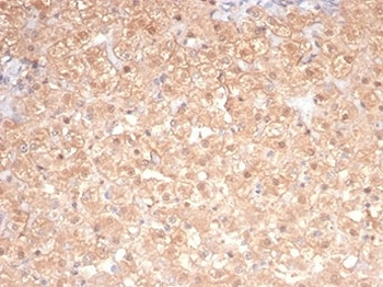
IHC staining of FFPE dog liver tissue with PRKCI antibody (clone PRKCI/4911). HIER: boil tissue sections in pH9 10mM Tris with 1mM EDTA for 20 min and allow to cool before testing.

IHC staining of FFPE cat liver tissue with PRKCI antibody (clone PRKCI/4911). HIER: boil tissue sections in pH9 10mM Tris with 1mM EDTA for 20 min and allow to cool before testing.

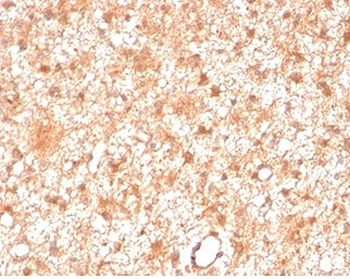
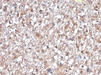
IHC staining of FFPE human brain tissue with PRKCI antibody (clone PRKCI/4911). HIER: boil tissue sections in pH9 10mM Tris with 1mM EDTA for 20 min and allow to cool before testing.

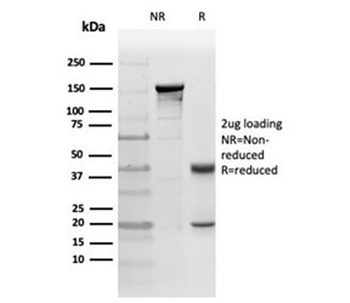
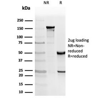
SDS-PAGE analysis of purified, BSA-free PRKCI antibody (clone PRKCI/4911) as confirmation of integrity and purity.

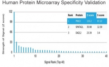
Analysis of HuProt (TM) microarray containing more than 19000 full-length human proteins using PRKCI antibody (clone PRKCI/4911). These results demonstrate the foremost specificity of the PRKCI/4911 mAb. Z- and S- score: The Z-score represents the strength of a signal that an antibody (in combination with a fluorescently-tagged anti-IgG secondary Ab) produces when binding to a particular protein on the HuProt (TM) array. Z-scores are described in units of standard deviations (SD's) above the mean value of all signals generated on that array. If the targets on the HuProt (TM) are arranged in descending order of the Z-score, the S-score is the difference (also in units of SD's) between the Z-scores. The S-score therefore represents the relative target specificity of an Ab to its intended target.
- Item 1 of 5
- Item 1 of 3
- Item 1 of 3
- Item 1 of 3