You have no items in your shopping cart.
Cart summary
Item 1 of 5
Item 1 of 5
PIN1 Antibody (Center)
Catalog Number: orb1928185

| Catalog Number | orb1928185 |
|---|---|
| Category | Antibodies |
| Description | Affinity Purified Rabbit Polyclonal Antibody (Pab) |
| Target | This PIN1 antibody is generated from rabbits immunized with a KLH conjugated synthetic peptide between 30-56 amino acids from the Central region of human PIN1. |
| Clonality | Polyclonal |
| Species/Host | Rabbit |
| Isotype | Rabbit IgG |
| Conjugation | Unconjugated |
| Reactivity | Human |
| Form/Appearance | Purified polyclonal antibody supplied in PBS with 0.09% (W/V) sodium azide. This antibody is purified through a protein A column, followed by peptide affinity purification. |
| UniProt ID | Q13526 |
| MW | 18243 Da |
| Tested applications | FC, IF, IHC-P, WB |
| Dilution range | IF: 1:10~50, WB: 1:1000, WB: 1:1000, IHC-P: 1:50~100, FC: 1:10~50 |
| Antibody Type | Primary Antibody |
| Clone Number | RB22881 |
| Storage | Maintain refrigerated at 2-8°C for up to 2 weeks. For long term storage store at -20°C in small aliquots to prevent freeze-thaw cycles |
| Alternative names | Peptidyl-prolyl cis-trans isomerase NIMA-interacti Read more... |
| Note | For research use only |
| NCBI | NP_006212.1 |
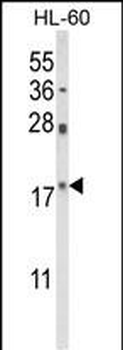
Western blot analysis of PIN1 Antibody (Center) in HL-60 cell line lysates (35 ug/lane). PIN1 (arrow) was detected using the purified Pab.
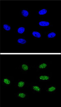
Confocal immunofluorescent analysis of PIN1 Antibody (Center) with 293 cell followed by Alexa Fluor 488-conjugated goat anti-rabbit lgG (green).DAPI was used to stain the cell nuclear (blue).
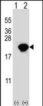
Western blot analysis of PIN1 (arrow) using rabbit polyclonal PIN1 Antibody (Center). 293 cell lysates (2 ug/lane) either nontransfected (Lane 1) or transiently transfected (Lane 2) with the PIN1 gene.
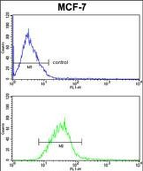
PIN1 Antibody (Center) FC analysis of MCF-7 cells (bottom histogram) compared to a negative control cell (top histogram). FITC-conjugated goat-anti-rabbit secondary antibodies were used for the analysis.
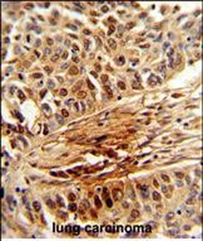
Formalin-fixed and paraffin-embedded human lung carcinoma reacted with PIN1 Antibody (Center), which was peroxidase-conjugated to the secondary antibody, followed by DAB staining. This data demonstrates the use of this antibody for immunohistochemistry; clinical relevance has not been evaluated.
