You have no items in your shopping cart.
Cart summary
Item 1 of 4
Item 1 of 4
PI3KCD Antibody / PI3K delta / p110delta
Catalog Number: orb1824480

| Catalog Number | orb1824480 |
|---|---|
| Category | Antibodies |
| Description | Phosphatidylinositol 3-kinase (PI 3-kinase) is composed of p85 and p110 subunits. p85 lacks PI 3-kinase activity and acts as an adapter, coupling p110 to activated protein tyrosine kinase. Two forms of p85 have been described (p85a and p85b), each possessing one SH3 and two SH2 domains. Various p110 forms have been identified. p110a and p110b interact with p85a, and p110a has also been shown to interact with p85b in vitro. It has been shown to bind p85a and b, but it apparently does not phosphorylate these subunits. p110d has the capacity to autophosphorylate and results in the nearly complete inactivation of the lipid kinase activity. Interestingly, p110g does not interact with the p85 subunits and has been shown to be activated by a and bg heterotrimeric G proteins. Two p110d isoforms have been identified and are widely expressed. Isoform 1 is expressed predominantly in leukocytes while isoform 2 is expressed in normal thymus, lung and spleen tissues. |
| Species/Host | Mouse |
| Clonality | Monoclonal |
| Clone Number | PIK3CD/4639 |
| Tested applications | IHC-P, WB |
| Reactivity | Human |
| Isotype | Mouse IgG2b, kappa |
| Immunogen | A recombinant fragment of human PIK3CD protein (within amino acids 520-720) was used as the immunogen for the PIK3CD antibody. |
| Antibody Type | Primary Antibody |
| Dilution range | Western blot: 1-2ug/ml,Immunohistochemistry (FFPE): 1-2ug/ml for 30 min at RT |
| Conjugation | Unconjugated |
| Formula | 0.2 mg/ml in 1X PBS with 0.1 mg/ml BSA (US sourced), 0.05% sodium azide |
| Hazard Information | This PIK3CD antibody is available for research use only. |
| UniProt ID | O00329 |
| Storage | Aliquot the PIK3CD antibody and store frozen at -20°C or colder. Avoid repeated freeze-thaw cycles. |
| Note | For research use only |
| Expiration Date | 12 months from date of receipt. |

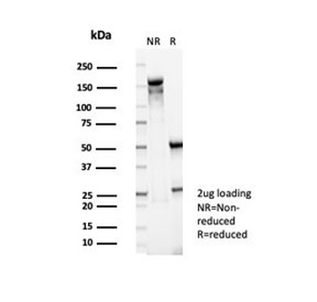
Western blot testing of human THP-1 cell lysate with PIK3CD antibody (clone PIK3CD/4639). Expected molecular weight: 110-120 kDa.

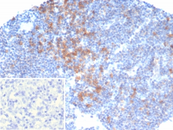
IHC staining of FFPE human lymph node tissue with PIK3CD antibody (clone PIK3CD/4639). Inset: PBS used in place of primary Ab (secondary Ab negative control). HIER: boil tissue sections in pH9 10 mM Tris with 1 mM EDTA for 20 min and allow to cool before testing.
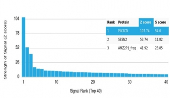
Analysis of a HuProt (TM) microarray containing more than 19000 full-length human proteins using PIK3R2 antibody (clone PIK3CD/4639). Z- and S- Score: The Z-score represents the strength of a signal that a monoclonal antibody (in combination with a fluorescently-tagged anti-IgG secondary antibody) produces when binding to a particular protein on the HuProt (TM) array. Z-scores are described in units of standard deviations (SD's) above the mean value of all signals generated on that array. If targets on HuProt (TM) are arranged in descending order of the Z-score, the S-score is the difference (also in units of SD's) between the Z-score. S-score therefore represents the relative target specificity of a mAb to its intended target. A mAb is considered to specific to its intended target, if the mAb has an S-score of at least 2.5. For example, if a mAb binds to protein X with a Z-score of 43 and to protein Y with a Z-score of 14, then the S-score for the binding of that mAb to protein X is equal to 29.

SDS-PAGE analysis of purified, BSA-free PIK3CD antibody (clone PIK3CD/4639) as confirmation of integrity and purity.
- Item 1 of 4
PI3KCD Antibody / PI3K delta / p110delta [orb1824479]
IHC-P, WB
Human
Mouse
Monoclonal
Unconjugated
100 μg - Item 1 of 4
PI3KCD Antibody / PI3K delta / p110delta [orb1824481]
IHC-P, WB
Human
Mouse
Monoclonal
Unconjugated
100 μg