You have no items in your shopping cart.
Cart summary
Item 1 of 3
Item 1 of 3
PDGF beta Antibody
Catalog Number: orb2636663

| Catalog Number | orb2636663 |
|---|---|
| Category | Antibodies |
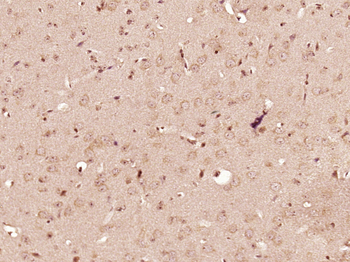
| Description | PDGF is a mitogen for mesenchyme- and glia-derived cells. It consists of two disulfide-bonded polypeptide chains, A and B, and occurs as three isoforms; PDGF AA, AB and BB. The three isoforms bind, with different affinities, to two receptor types, receptors. Evidence suggests that PDGF may function as a neurotrophic factor. Receptors for PDGF-A are expressed in oligodendrocyte progenitor cells whereas receptors for PDGF-B are expressed on neurons. These facts suggest that the different isoforms of PDGF may regulate growth and differentiation of different cell types in the developing central nervous system through paracrine and autocrine routes. |
| Species/Host | Mouse |
| Clonality | Monoclonal |
| Clone Number | PDGFB/3072 |
| Tested applications | IHC-P |
| Reactivity | Human |
| Isotype | Mouse IgG2c, kappa |
| Immunogen | A portion of amino acids 27-158 was used as the immunogen for the PDGFB antibody. |
| Antibody Type | Primary Antibody |
| Dilution range | Immunohistochemistry (FFPE): 1-2ug/ml |
| Purity | Protein A/G affinity |
| Conjugation | Unconjugated |
| Formula | 0.2 mg/ml in 1X PBS with 0.1 mg/ml BSA (US sourced), 0.05% sodium azide |
| Hazard Information | This PDGFB antibody is available for research use only. |
| UniProt ID | P01127 |
| Storage | Maintain refrigerated at 2-8°C for up to 2 weeks. For long term storage store at -20°C in small aliquots to prevent freeze-thaw cycles. |
| Buffer/Preservatives | 0.2 mg/ml in 1X PBS with 0.1 mg/ml rAlbumin (US sourced), 0.05% sodium azide |
| Note | For research use only |
| Application notes | Optimal dilution of the PDGFB antibody should be determined by the researcher. |
| Expiration Date | 12 months from date of receipt. |

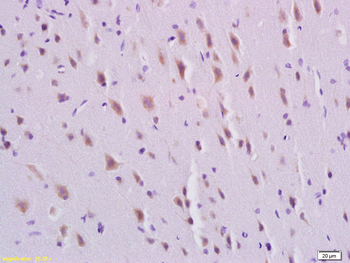
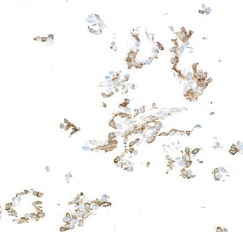
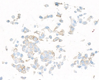
Analysis of HuProt (TM) microarray containing more than 19000 full-length human proteins using PDGFB antibody (clone PDGFB/3072). These results demonstrate the foremost specificity of the PDGFB/3072 mAb. Z- and S- score: The Z-score represents the strength of a signal that an antibody (in combination with a fluorescently-tagged anti-IgG secondary Ab) produces when binding to a particular protein on the HuProt (TM) array. Z-scores are described in units of standard deviations (SD's) above the mean value of all signals generated on that array. If the targets on the HuProt (TM) are arranged in descending order of the Z-score, the S-score is the difference (also in units of SD's) between the Z-scores. The S-score therefore represents the relative target specificity of an Ab to its intended target.
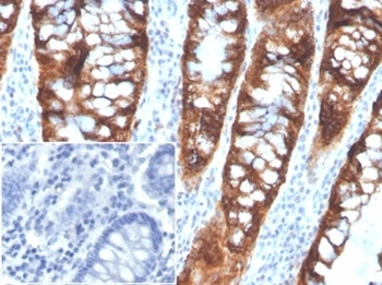
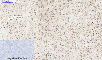
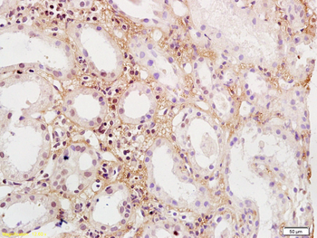
IHC staining of FFPE human colon tissue with PDGFB antibody (clone PDGFB/3072). Negative control inset: PBS instead of primary antibody to control for secondary binding. HIER: boil tissue sections in pH9 10mM Tris with 1mM EDTA for 20 min and allow to cool before testing.
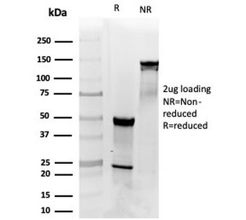
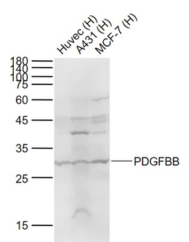
SDS-PAGE analysis of purified, BSA-free PDGFB antibody (clone PDGFB/3072) as confirmation of integrity and purity.
- Item 1 of 7
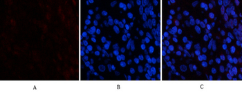
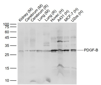
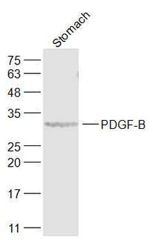
PDGF-B Polyclonal Antibody [orb1412572]
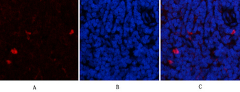
IF, IHC-P, WB
Human, Mouse, Rat
Rabbit
Polyclonal
Unconjugated
100 μl - Item 1 of 4
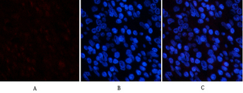
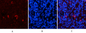
PDGFBB Rabbit Polyclonal Antibody [orb11255]
IF, IHC-Fr, IHC-P, WB
Mouse, Rat
Human, Mouse, Rat
Rabbit
Polyclonal
Unconjugated
50 μl, 100 μl, 200 μl - Item 1 of 3
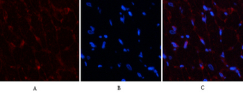
PDGF-B Rabbit Polyclonal Antibody [orb11254]
FC, IF, IHC-Fr, IHC-P, WB
Bovine, Canine, Equine, Rabbit, Sheep
Human, Mouse, Rat
Rabbit
Polyclonal
Unconjugated
100 μl, 200 μl, 50 μl - Item 1 of 3
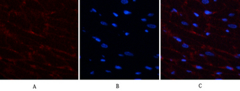
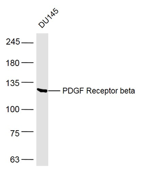
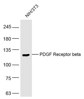
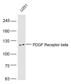
PDGF Receptor beta Rabbit Polyclonal Antibody [orb11257]
IF, IHC-Fr, IHC-P, WB
Bovine, Canine, Mouse
Human, Rat
Rabbit
Polyclonal
Unconjugated
200 μl, 100 μl, 50 μl - Item 1 of 5
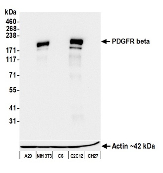
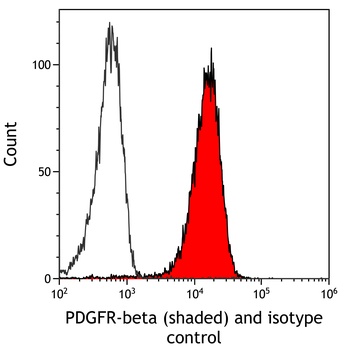
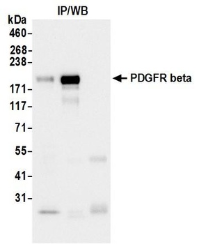
Rabbit anti-PDGFR beta Recombinant Monoclonal Antibody [orb1570942]
FC, ICC, IP, WB
Mouse
Rabbit
Recombinant
Unconjugated
10 μl