You have no items in your shopping cart.
Cart summary
Item 1 of 5
Item 1 of 5
PAX6-T373 Antibody
Catalog Number: orb1263581

| Catalog Number | orb1263581 |
|---|---|
| Category | Antibodies |
| Description | PAX6-T373 Antibody |
| Target | PAX6 |
| Clonality | Polyclonal |
| Isotype | Rabbit Ig |
| Conjugation | Unconjugated |
| Reactivity | Human, Rat |
| Form/Appearance | Liquid |
| Concentration | batch dependent |
| Buffer/Preservatives | Supplied in PBS with 0.09% (W/V) sodium azide. |
| Immunogen | This PAX6 antibody is generated from rabbits immunized with a KLH conjugated synthetic peptide between 352-380 amino acids from human PAX6. |
| UniProt ID | P26367 |
| MW | 47 kDa |
| Tested applications | IF, IHC-P, WB |
| Application notes | For WB starting dilution is: 1:1000For IHC-P starting dilution is: 1:10~50For IF starting dilution is: 1:10~50 |
| Antibody Type | Primary Antibody |
| Storage | Maintain refrigerated at 2-8°C for up to 2 weeks. For long term storage store at -20°C in small aliquots to prevent freeze-thaw cycles. |
| Alternative names | Paired box protein Pax-6, Aniridia type II protein Read more... |
| Note | For research use only |
| NCBI | P26367 |
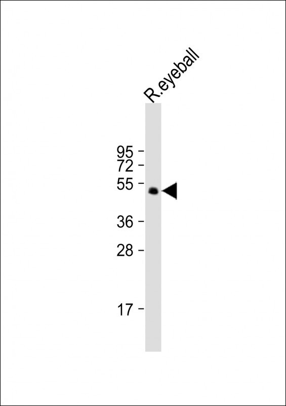
Western Blot at 1:2000 dilution + rat eyeball lysate Lysates/proteins at 20 ug per lane.
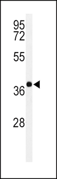
Western blot analysis of PAX6-T373 in Y79 cell line lysates (35 ug/lane)
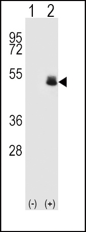
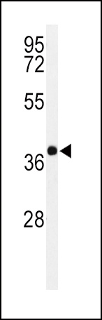
Western blot analysis of PAX6 using rabbit polyclonal PAX6 Antibody (T373) using 293 cell lysates (2 ug/lane) either nontransfected (Lane 1) or transiently transfected (Lane 2) with the PAX6 gene.
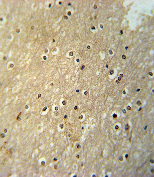
PAX6-T373 Antibody immunohistochemistry analysis in formalin fixed and paraffin embedded human brain tissue followed by peroxidase conjugation of the secondary antibody and DAB staining.
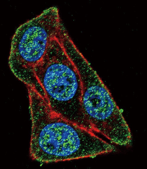
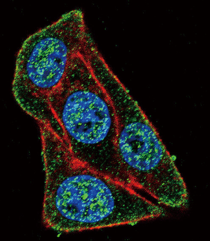
Confocal immunofluorescent analysis of PAX6-T373 Antibody with Hela cell followed by Alexa Fluor 488-conjugated goat anti-rabbit lgG (green). Actin filaments have been labeled with Alexa Fluor 555 phalloidin (red).DAPI was used to stain the cell nuclear (blue).
- Item 1 of 5