You have no items in your shopping cart.
Cart summary
Item 1 of 4
Item 1 of 4
p63 Antibody / Tumor protein 63
Catalog Number: orb2636611

| Catalog Number | orb2636611 |
|---|---|
| Category | Antibodies |
| Description | p63 is a homolog of the tumor suppressor p53. It is identified in basal cells in the epithelial layers of a variety of tissues, including epidermis, cervix, urothelium, breast and prostate. p63 was detected in nuclei of the basal epithelium in normal prostate glands; however, it was not expressed in malignant tumors of the prostate. As a result, p63 has been reported as a useful marker for differentiating benign from malignant lesions in the prostate, particularly when used in combination with markers of high molecular weight cytokeratins and the prostate-specific marker AMACR (P504S). p63 has also been shown to be a sensitive marker for lung squamous cell carcinomas (SqCC), with a sensitivity of ~90%. Specificity for lung SqCC, vs. lung adenocarcinoma (LADC), is approximately 80%. In breast tissue, p63 has been identified in myoepithelial cells of normal ducts. |
| Clonality | Recombinant |
| Species/Host | Rabbit |
| Isotype | Rabbit IgG, kappa |
| Conjugation | Unconjugated |
| Reactivity | Human |
| Buffer/Preservatives | 0.2 mg/ml in 1X PBS with 0.1 mg/ml rAlbumin (US sourced), 0.05% sodium azide |
| Purity | Protein A affinity |
| Immunogen | A portion of amino acids 600-680 was used as the immunogen for the recombinant p63 antibody. |
| UniProt ID | Q9H3D4 |
| Tested applications | IHC-P |
| Dilution range | Immunohistochemistry (FFPE): 1-2ug/ml |
| Application notes | Optimal dilution of the recombinant p63 antibody should be determined by the researcher. |
| Antibody Type | Primary Antibody |
| Clone Number | TP63/4379R |
| Formula | 0.2 mg/ml in 1X PBS with 0.1 mg/ml BSA (US sourced), 0.05% sodium azide |
| Storage | Maintain refrigerated at 2-8°C for up to 2 weeks. For long term storage store at -20°C in small aliquots to prevent freeze-thaw cycles. |
| Hazard Information | This recombinant p63 antibody is available for research use only. |
| Note | For research use only |

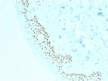
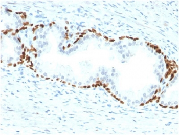
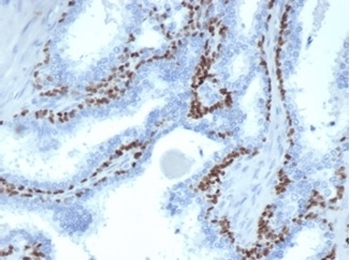
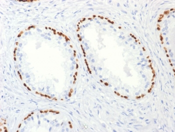
IHC staining of FFPE human prostate cancer with recombinant p63 antibody (clone TP63/4379R). HIER: boil tissue sections in pH9 10mM Tris with 1mM EDTA for 20 min and allow to cool before testing.
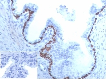
IHC staining of FFPE human prostate cancer tissue with recombinant p63 antibody (clone TP63/4379R). Negative control inset: PBS instead of primary antibody to control for secondary binding. HIER: boil tissue sections in pH9 10mM Tris with 1mM EDTA for 20 min and allow to cool before testing.
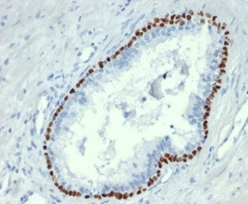
IHC staining of FFPE human prostate cancer tissue with recombinant p63 antibody (clone TP63/4379R). HIER: boil tissue sections in pH9 10mM Tris with 1mM EDTA for 20 min and allow to cool before testing.

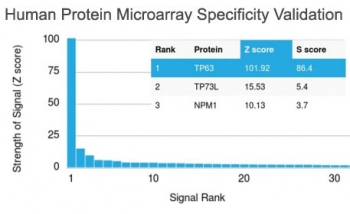
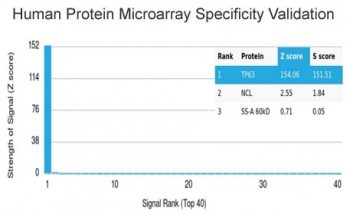
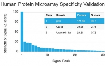
Analysis of HuProt (TM) microarray containing more than 19000 full-length human proteins using recombinant p63 antibody (clone TP63/4379R). These results demonstrate the foremost specificity of the TP63/4379R mAb. Z- and S- score: The Z-score represents the strength of a signal that an antibody (in combination with a fluorescently-tagged anti-IgG secondary Ab) produces when binding to a particular protein on the HuProt (TM) array. Z-scores are described in units of standard deviations (SD's) above the mean value of all signals generated on that array. If the targets on the HuProt (TM) are arranged in descending order of the Z-score, the S-score is the difference (also in units of SD's) between the Z-scores. The S-score therefore represents the relative target specificity of an Ab to its intended target.
- Item 1 of 5
- Item 1 of 5
- Item 1 of 4
- Item 1 of 3
- Item 1 of 3