You have no items in your shopping cart.
Cart summary
Item 1 of 4
Item 1 of 4
NeuroD1 Antibody
Catalog Number: orb1265129

| Catalog Number | orb1265129 |
|---|---|
| Category | Antibodies |
| Description | NeuroD1 Antibody |
| Species/Host | Rabbit |
| Clonality | Polyclonal |
| Tested applications | IF, IHC-P, WB |
| Reactivity | Human |
| Isotype | Rabbit Ig |
| Immunogen | This NeuroD1 antibody is generated from rabbits immunized with a KLH conjugated synthetic peptide between 15-45 amino acids from the N-terminal region of human NeuroD1. |
| Antibody Type | Primary Antibody |
| Concentration | batch dependent |
| Form/Appearance | Liquid |
| Conjugation | Unconjugated |
| MW | 40 kDa |
| Target | NEUROD1 |
| UniProt ID | Q13562 |
| NCBI | Q13562 |
| Storage | Maintain refrigerated at 2-8°C for up to 2 weeks. For long term storage store at -20°C in small aliquots to prevent freeze-thaw cycles. |
| Buffer/Preservatives | Supplied in PBS with 0.09% (W/V) sodium azide. |
| Alternative names | Neurogenic differentiation factor 1, NeuroD, Neuro Read more... |
| Note | For research use only |
| Application notes | For WB starting dilution is: 1:1000For IF starting dilution is: 1:10~50For IHC-P starting dilution is: 1:50~100 |
| Expiration Date | 12 months from date of receipt. |
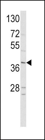
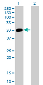
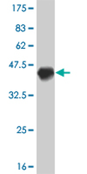
Western blot analysis of hNeuroD1-Q30 in HepG2 cell line lysates (35 ug/lane)
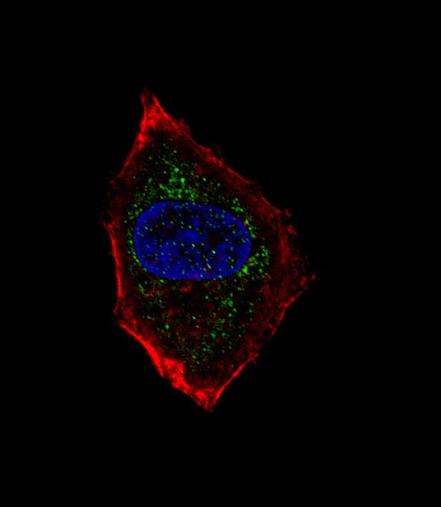
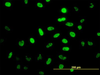
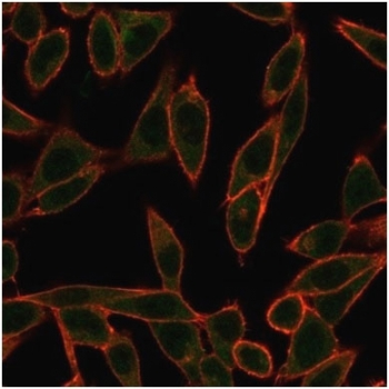
Fluorescent confocal image of HepG2 cell stained with hNeuroD1-Q30. HepG2 cells were fixed with 4% PFA (20 min), permeabilized with Triton X-100 (0.1%, 10 min), then incubated with hNeuroD1-Q30 primary antibody (1:25). For secondary antibody, Alexa Fluor 488 conjugated donkey anti-rabbit antibody (green) was used (1:400). Cytoplasmic actin was counterstained with Alexa Fluor 555 (red) conjugated Phalloidin (7 units/ml). Nuclei were counterstained with DAPI (blue) (10 ug/ml, 10 min). hNeuroD1-Q30 immunoreactivity is localized to vesicles significantly.
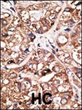
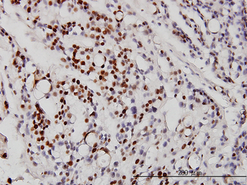
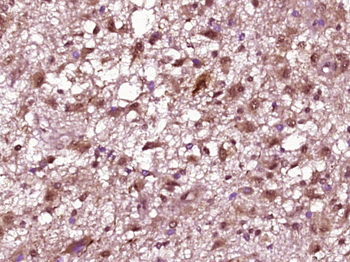
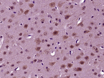
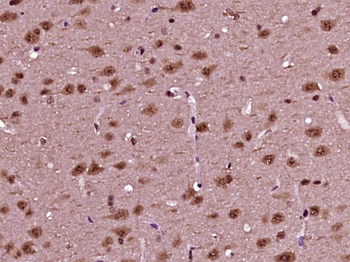
Formalin-fixed and paraffin-embedded human cancer tissue reacted with the primary antibody, which was peroxidase-conjugated to the secondary antibody, followed by DAB staining. BC = breast carcinoma; HC = hepatocarcinoma.
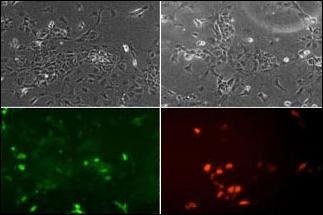
ES cells were transiently transfected with flag-tagged mouse NeuroD1 (tagged on N-term).Fixed 24h post transfection.Stained for flag tag (red) to check that some cells express protein. Most protein was in nucleus but some was cytoplasmic. Stained with NeuroD1 N-term antibodies at 1:100. NeuroD1 N-term antibody showed strong and clear staining with similar pattern to the flag staining. (Supplied by Sally Lowell, Edinburgh University)
- Item 1 of 5
NEUROD1 monoclonal antibody (M01), clone 3H8 [orb2292520]
ELISA, IF, IHC-P, WB
Human
Mouse
Monoclonal
Unconjugated
100 μg - Item 1 of 4
- Item 1 of 4
- Item 1 of 4
- Item 1 of 4
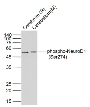
Phospho-NEUROD1 (Ser274) Rabbit Polyclonal Antibody [orb221716]
IF, IHC-Fr, IHC-P, WB
Bovine, Porcine, Sheep
Human, Mouse, Rat
Rabbit
Polyclonal
Unconjugated
50 μl, 100 μl, 200 μl