You have no items in your shopping cart.
Cart summary
Item 1 of 4
Item 1 of 4
NDUFB4 Antibody
Catalog Number: orb1265007

| Catalog Number | orb1265007 |
|---|---|
| Category | Antibodies |
| Description | NDUFB4 Antibody |
| Species/Host | Rabbit |
| Clonality | Polyclonal |
| Tested applications | FC, IF, IHC-P, WB |
| Reactivity | Human |
| Isotype | Rabbit Ig |
| Immunogen | This NDUFB4 antibody is generated from a rabbit immunized with a KLH conjugated synthetic peptide between 3-36 amino acids from the N-terminal region of human NDUFB4. |
| Concentration | batch dependent |
| Dilution range | For IHC-P starting dilution is: 1:25For IF starting dilution is: 1:25For FACS starting dilution is: 1:25For WB starting dilution is: 1:1000 |
| Form/Appearance | Liquid |
| Conjugation | Unconjugated |
| MW | 15 kDa |
| Target | NDUFB4 |
| UniProt ID | O95168 |
| NCBI | O95168 |
| Storage | Store at 4°C for three months and -20°C, stable for up to one year. As with all antibodies care should be taken to avoid repeated freeze thaw cycles. Antibodies should not be exposed to prolonged high temperatures. |
| Buffer/Preservatives | Supplied in PBS with 0.09% (W/V) sodium azide. |
| Alternative names | NADH dehydrogenase [ubiquinone] 1 beta subcomplex Read more... |
| Note | For research use only |
| Application notes | For IHC-P starting dilution is: 1:25For IF starting dilution is: 1:25For FACS starting dilution is: 1:25For WB starting dilution is: 1:1000 |
| Expiration Date | 12 months from date of receipt. |
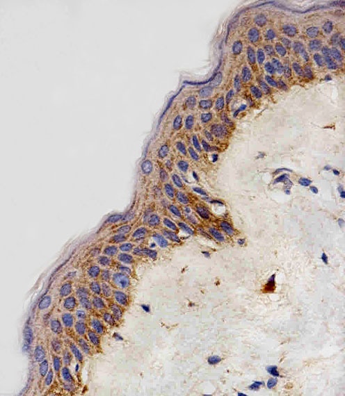
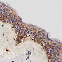
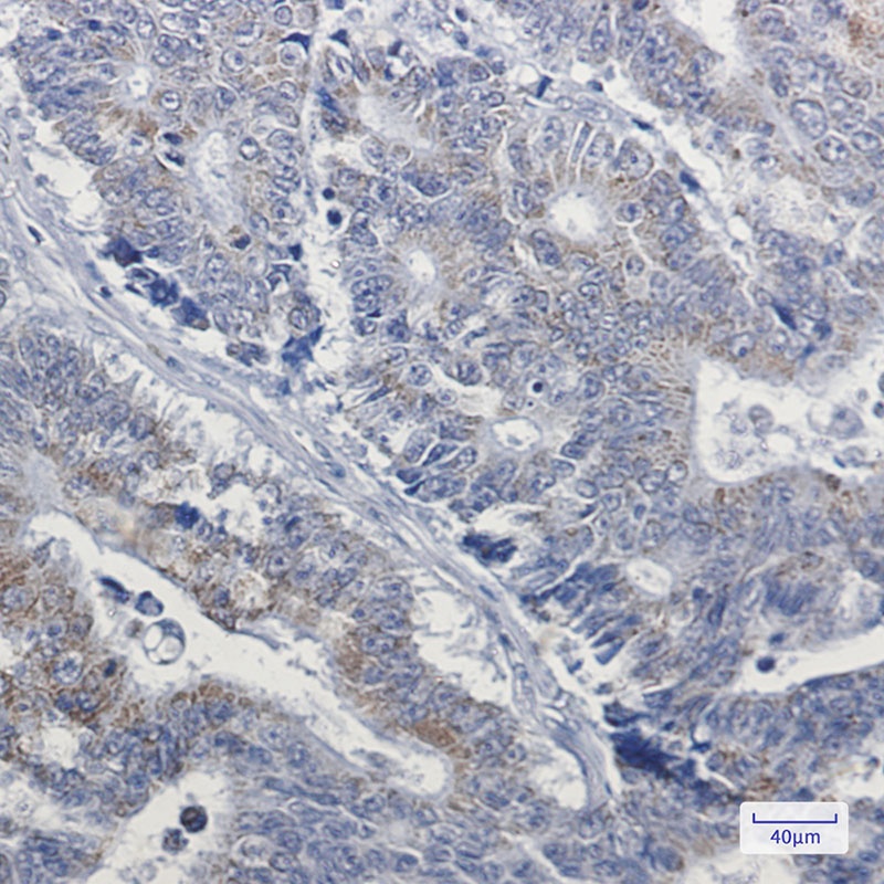
Immunohistochemical analysis of paraffin-embedded H. skin section using NDUFB4 Antibody (N-term). Antibody was diluted at 1:100 dilution. A peroxidase-conjugated goat anti-rabbit IgG at 1:400 dilution was used as the secondary antibody, followed by DAB staining.
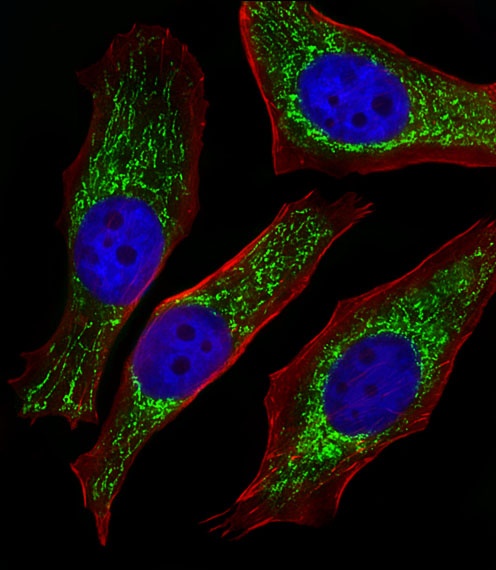
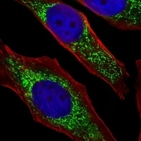
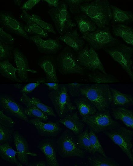
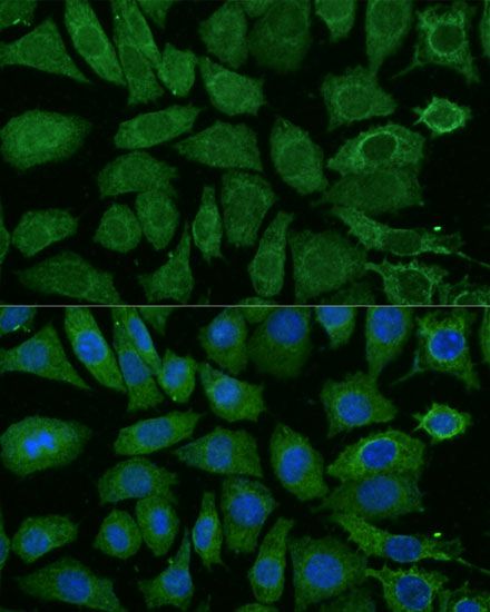
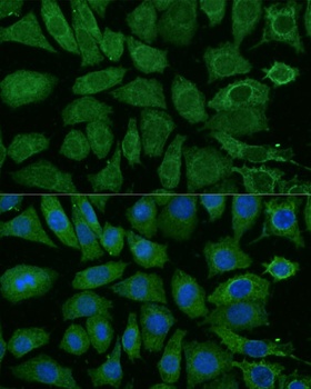
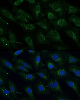
Fluorescent image of Hela cells stained with NDUFB4 Antibody (N-term). Antibody was diluted at 1:25 dilution. An Alexa Fluor 488-conjugated goat anti-rabbit lgG at 1:400 dilution was used as the secondary antibody (green). DAPI was used to stain the cell nuclear (blue). Cytoplasmic actin was counterstained with Alexa Fluor 555 conjugated with Phalloidin (red).
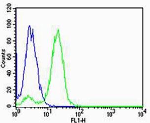
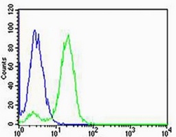
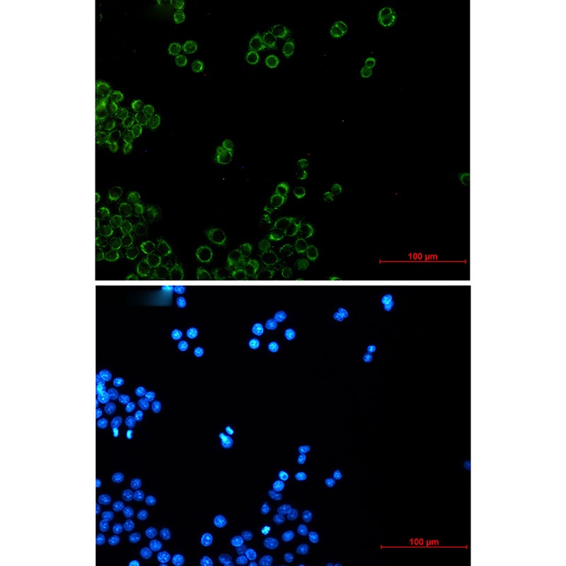
Flow cytometric analysis of Hela cells using NDUFB4 Antibody (N-term) (green) compared to an isotype control of rabbit IgG (blue). Antibody was diluted at 1:25 dilution. An Alexa Fluor 488 goat anti-rabbit lgG at 1:400 dilution was used as the secondary antibody.
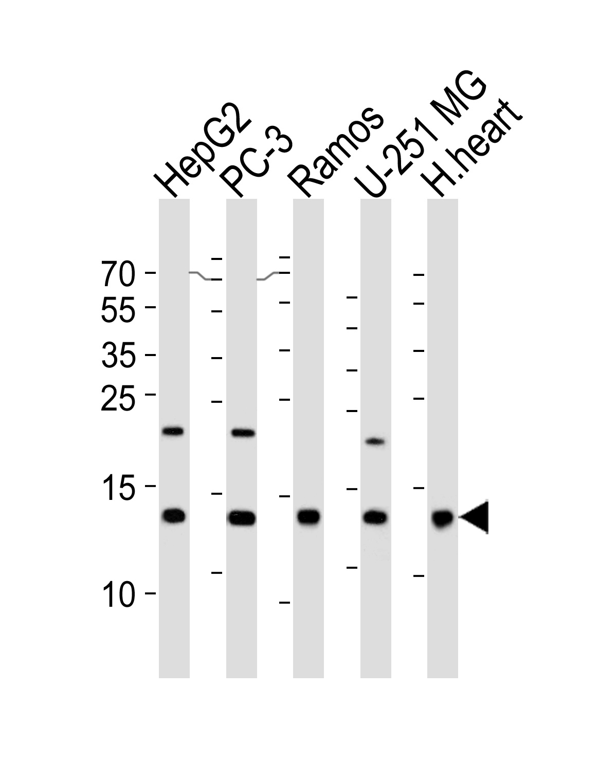
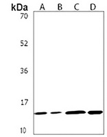
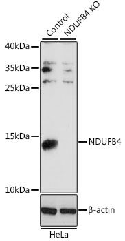
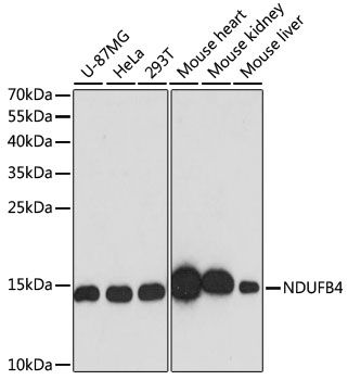
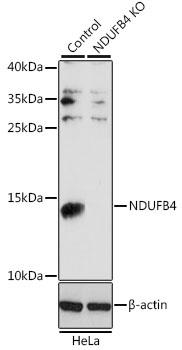
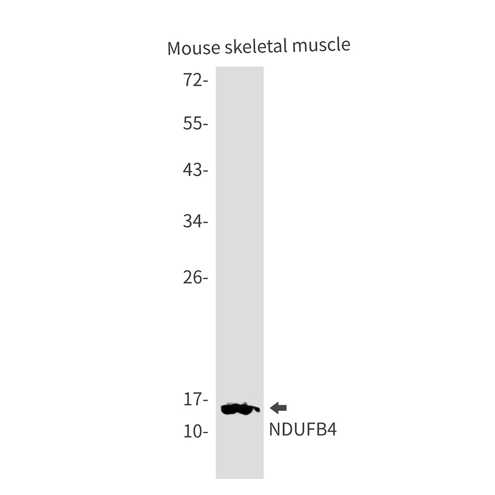
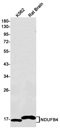
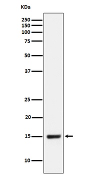
Western blot analysis of lysates from HepG2, PC-3, Ramos, U-251 MG cell line and human heart tissue lusyte (from left to right), using NDUFB4 Antibody at 1:1000 at each lane.
- Item 1 of 4
- Item 1 of 4
NDUFB4 antibody [orb540601]
ICC, IF, IHC, WB
Human, Mouse, Rat
Polyclonal
Unconjugated
200 μl, 100 μl, 50 μl - Item 1 of 4
- Item 1 of 4
NDUFB4 Antibody [orb1564560]
ICC, IHC-Fr, IHC-P, WB
Human, Mouse, Rat
Rabbit
Monoclonal
Unconjugated
100 μl, 50 μl, 20 μl - Item 1 of 1
NDUFB4 Rabbit Monoclonal Antibody [orb1147272]
ICC, IF, IHC, WB
Human, Mouse, Rat
Rabbit
Monoclonal
Unconjugated
30 μl, 100 μl

Submit a review
Filter by Rating

- 5 stars

- 4 stars
- 3 stars
- 2 stars
- 1 stars
