You have no items in your shopping cart.
Description
Images & Validation
−Item 1 of 4
| Tested Applications | FLISA, IF, WB |
|---|---|
| Dilution Range | FLISA: 1:10,000 - 1:50,000, IF: 1:500 - 1:2,500 |
| Reactivity | Mouse |
| Application Notes |
Key Properties
−| Antibody Type | Secondary Antibody |
|---|---|
| Host | Rabbit |
| Clonality | Polyclonal |
| Isotype | IgG |
| Immunogen | Mouse IgG1 heavy chain |
| Purity | This product was prepared from monospecific antiserum by immunoaffinity chromatography using antigens coupled to agarose beads followed by solid phase adsorption(s) to remove any unwanted reactivities. Assay by immunoelectrophoresis resulted in a single precipitin arc against anti-Fluorescein, anti-Rabbit Serum, Mouse IgG and Mouse Serum. Specificity was confirmed by ELISA. |
| Conjugation | FITC |
Storage & Handling
−| Storage | Store secondary antibody at 4° C prior to restoration. For extended storage aliquot contents and freeze at -20° C or below. Avoid cycles of freezing and thawing. Centrifuge product if not completely clear after standing at room temperature. This product is stable for several weeks at 4° C as an undiluted liquid. Dilute only prior to immediate use. |
|---|---|
| Form/Appearance | Lyophilized |
| Buffer/Preservatives | Preservative: 0.01% (w/v) Sodium Azide. Stabilizer: 10 mg/mL Bovine Serum Albumin (rAlbumin) - Immunoglobulin and Protease free; Buffer: 0.02 M Potassium Phosphate, 0.15 M Sodium Chloride, pH 7.2 |
| Concentration | 1.0 mg/mL |
| Expiration Date | 12 months from date of receipt. |
| Disclaimer | For research use only |
Alternative Names
−Rabbit Anti-Mouse IgG1 (Gamma 1 chain) Antibody fluorescein Conjugation, Rabbit Anti-Mouse IgG1 FITC Conjugated Antibody
Quality Guarantee
Explore bioreagents carefree to elevate your research. All our products are rigorously tested for performance. If a product does not perform as described on its datasheet, our scientific support team will provide expert troubleshooting, a prompt replacement, or a refund. For full details, please see our Terms & Conditions and Buying Guide. Contact us at [email protected].
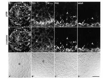
Colocalization of p120 and β-catenin using Anti-Mouse IgG1 FITC (p/n orb347540) during postnatal testis development. Here, the 8D11 antibody was used to immunostain for p120. In all locations, p120 and β-catenin showed exact colocalization. At Postnatal Day 7 (A), diffuse immunostaining outlined all cells of the developing epithelium (solid arrow). In addition, punctate structures (arrowhead) were observed throughout the tubule. At the tubule periphery, intense immunostaining was associated with peritubular cells (dashed arrows). From Day 21 through adulthood, p120 and β-catenin colocalized at basal inter-Sertoli junctions (arrows in B–D), and at punctate, spermatocyte-associated structures (arrowheads in B–D). In addition, extended, linear immunostaining at the level of spermatocytes was observed at Day 43 (C and C'; dashed arrows). Corresponding DIC images are shown in A", B", C", and D". The center (C) of Day 7 and Day 21 seminiferous tubules is indicated in the DIC images. In all images, the seminiferous tubule basement membrane is located at the bottom, and in C" and D", the entire seminiferous epithelium is shown. Bar = 20 µm.
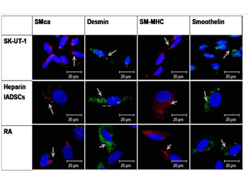
Isolated hADSCs (IADSCs) were differentiated into SMCs using retinoic acid (RA), heparin was used as a positive control, while SK-UT-1 cells was used as a SMC control. Expression of SMC markers smooth muscle alpha actin (SM-αa, red, Texas Red Conjugated anti-Mouse IgG2, γ2a chain specific), desmin (green, Fluorescein Conjugated anti-Mouse IgG1, γ1 chain specific), smooth muscle myosin heavy chain (SM-MHC, red, Texas Red Conjugated anti-Mouse κ, kappa chain specific), and smoothelin (green, Fluorescein Conjugated anti-Mouse IgG1, γ1 chain specific) in differentiated SMCs was determined by indirect immunofluorescence. Nuclei were counter stained with DAPI (blue). Expression of all four markers can be seen in all the cells, particularly in RA differentiated SMCs.
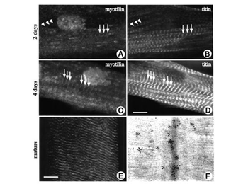
Localization of myotilin in differentiating and mature myocytes. Human skeletal muscle cells were grown on glass coverslips, differentiated for 4 days and stained with myotilin antibody (A and C) and with Z-disc specific titin antibody T12 mAb IgG1 (B and D) using Anti-Mouse IgG1 FITC. During the early stages of myofibril assembly, myotilin expression was faint and diffuse. The protein was concentrated at the areas where myofibrils are formed, and, although numerous Z-discs could be discerned, only very few of them contained myotilin (arrows in A and B). Only at the stage of myofibril alignment did myotilin appear at the mature, aligned Z-discs (arrows in C and D). In mature human skeletal muscle sections myotilin is found in a cross-striated pattern (E). Immunoelectron microscopic analysis of mature human skeletal muscle stained with myotilin antibody reveals a decoration of Z-discs (F). Bar = 10 µm.
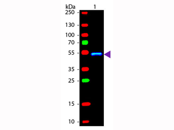
Western blot of Fluorescein conjugated Rabbit Anti-Mouse IgG1 (Gamma 1 chain) secondary antibody. Lane 1: Mouse IgG1. Lane 2: None. Load: 50 ng per lane. Primary antibody: None. Secondary antibody: Fluorescein rabbit secondary antibody at 1:1000 for 60 min at RT. Blocking: orb348637 for 30 min at RT. Predicted/Observed size: 55 kDa, 55 kDa for Mouse IgG1 (Gamma 1 chain). Other band(s): None.
Documents Download
Datasheet
Product Information
Request a Document
Protocol Information
WB
Western Blot (IB, immunoblot)
IF
Immunofluorescence
Mouse IgG1 Secondary Antibody Fluorescein Conjugated (orb347540)

- 0.0
Based on 0 reviews
Participating in our Biorbyt product reviews program enables you to support fellow scientists by sharing your firsthand experience with our products.
Login to Submit a ReviewAvailable Sizes
Select a size below
Free Secondary Antibody (20 ul)0/0
Please add an antibody product to your cart first.