You have no items in your shopping cart.
Cart summary
Item 1 of 4
Item 1 of 4
MLX Antibody (Center)
Catalog Number: orb1928379

| Catalog Number | orb1928379 |
|---|---|
| Category | Antibodies |
| Description | Affinity Purified Rabbit Polyclonal Antibody (Pab) |
| Species/Host | Rabbit |
| Clonality | Polyclonal |
| Clone Number | RB22641 |
| Tested applications | FC, IHC-P, WB |
| Predicted Reactivity | Mouse |
| Reactivity | Human |
| Isotype | Rabbit IgG |
| Antibody Type | Primary Antibody |
| Dilution range | WB: 1:1000, WB: 1:1000, IHC-P: 1:50~100, FC: 1:10~50 |
| Form/Appearance | Purified polyclonal antibody supplied in PBS with 0.09% (W/V) sodium azide. This antibody is purified through a protein A column, followed by peptide affinity purification. |
| Conjugation | Unconjugated |
| MW | 33300 Da |
| Target | This MLX antibody is generated from rabbits immunized with a KLH conjugated synthetic peptide between 125-151 amino acids from the Central region of human MLX. |
| UniProt ID | Q9UH92 |
| NCBI | NP_937848.1, NP_937847.1, NP_733752.1 |
| Storage | Maintain refrigerated at 2-8°C for up to 2 weeks. For long term storage store at -20°C in small aliquots to prevent freeze-thaw cycles |
| Alternative names | Max-like protein X, Class D basic helix-loop-helix Read more... |
| Note | For research use only |
| Expiration Date | 12 months from date of receipt. |
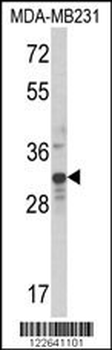
Western blot analysis of MLX Antibody (Center) in MDA-MB231 cell line lysates (35 ug/lane). MLX (arrow) was detected using the purified Pab.
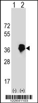
Western blot analysis of MLX (arrow) using rabbit polyclonal MLX Antibody (Center). 293 cell lysates (2 ug/lane) either nontransfected (Lane 1) or transiently transfected (Lane 2) with the MLX gene.
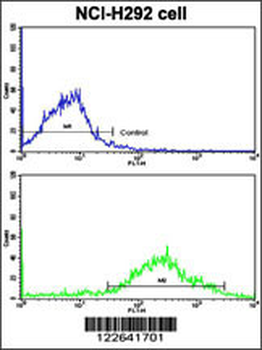
Flow cytometric analysis of NCI-H292 cells using MLX Antibody (Center) (bottom histogram) compared to a negative control cell (top histogram). FITC-conjugated goat-anti-rabbit secondary antibodies were used for the analysis.
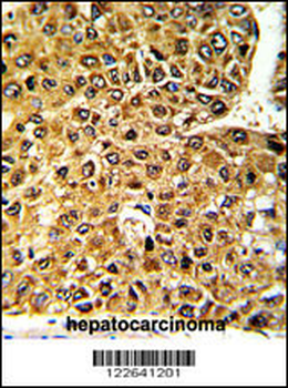
Formalin-fixed and paraffin-embedded human hepatocarcinoma with MLX Antibody (Center), which was peroxidase-conjugated to the secondary antibody, followed by DAB staining. This data demonstrates the use of this antibody for immunohistochemistry; clinical relevance has not been evaluated.
