You have no items in your shopping cart.
Cart summary
Item 1 of 5
Item 1 of 5
MIF Antibody / Macrophage migration inhibitory factor
Catalog Number: orb1824113

| Catalog Number | orb1824113 |
|---|---|
| Category | Antibodies |
| Description | Macrophage migration inhibitory factor, known as MIF or glycosylation inhibiting factor, is a secreted, homotrimeric, pro-inflammatory cytokine that modulates macrophage and T cell function and is an important regulator of host response to infection. MIF is expressed at sites of inflammation, which suggests that it plays a role in regulating macrophage function in host defense. MIF is produced by the pituitary gland and is found in monocytes, macrophages, differentiating immunological cells in the eye lens and brain, and fibroblasts. Elevated levels of MIF protein are detected in the plasma of patients with severe sepsis or septic shock, a condition where MIF influences endotoxic shock by enhancing the production of other inflammatory cytokines including tumor necrosis factor Alpha (TNF-Alpha), interleukin-1 (IL-1) and interferon-Gamma (IFN-Gamma). MIF promotes the systemic inflammatory response by counter-regulating glucocorticoid-mediated inhibition of immune-cell activation and proinflammatory cytokine production. MIF may mediate tissue destruction through the induction of proteinases. |
| Species/Host | Mouse |
| Clonality | Monoclonal |
| Clone Number | MIF/6280 |
| Tested applications | IHC-P, WB |
| Reactivity | Human |
| Isotype | Mouse IgG |
| Immunogen | A recombinant fragment of human protein was used as the immunogen for the MIF antibody. |
| Antibody Type | Primary Antibody |
| Dilution range | Western blot: 1-2ug/ml,Immunohistochemistry (FFPE): 1-2ug/ml for 30 min at RT |
| Conjugation | Unconjugated |
| Formula | 1 mg/ml in 1X PBS; BSA free, sodium azide free |
| Hazard Information | This MIF antibody is available for research use only. |
| UniProt ID | P14174 |
| Storage | Aliquot the MIF antibody and store frozen at -20°C or colder. Avoid repeated freeze-thaw cycles. |
| Note | For research use only |
| Expiration Date | 12 months from date of receipt. |

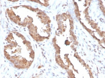
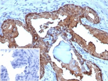
IHC staining of FFPE human prostate tissue with MIF antibody (clone MIF/6280). HIER: boil tissue sections in pH9 10 mM Tris with 1 mM EDTA for 20 min and allow to cool before testing.
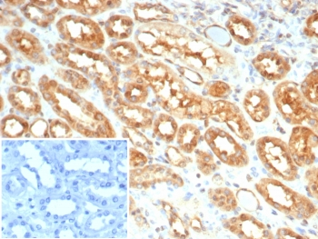
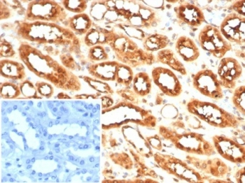
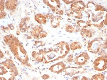
IHC staining of FFPE human kidney tissue with MIF antibody (clone MIF/6280). Inset: PBS used in place of primary Ab (secondary Ab negative control). HIER: boil tissue sections in pH9 10 mM Tris with 1 mM EDTA for 20 min and allow to cool before testing.
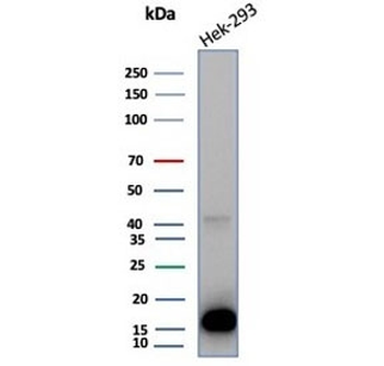
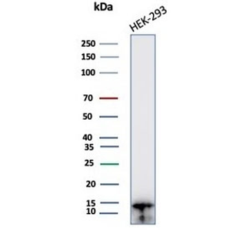
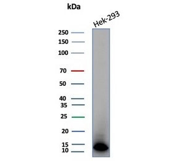
Western blot testing of human HEK293 cell lysate with MIF antibody (clone MIF/6280). Predicted molecular weight ~13 kDa.
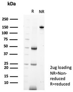
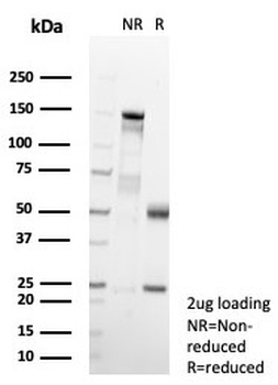
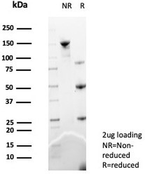
SDS-PAGE analysis of purified, BSA-free MIF antibody (clone MIF/6280) as confirmation of integrity and purity.
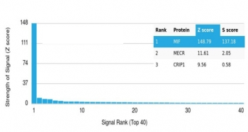
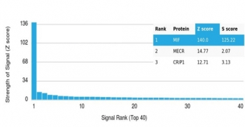
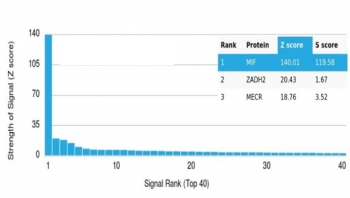
Analysis of a HuProt (TM) microarray containing more than 19000 full-length human proteins using MIF antibody (clone MIF/6280). Z- and S- Score: The Z-score represents the strength of a signal that a monoclonal antibody (in combination with a fluorescently-tagged anti-IgG secondary antibody) produces when binding to a particular protein on the HuProt (TM) array. Z-scores are described in units of standard deviations (SD's) above the mean value of all signals generated on that array. If targets on HuProt (TM) are arranged in descending order of the Z-score, the S-score is the difference (also in units of SD's) between the Z-score. S-score therefore represents the relative target specificity of a mAb to its intended target. A mAb is considered to specific to its intended target, if the mAb has an S-score of at least 2.5. For example, if a mAb binds to protein X with a Z-score of 43 and to protein Y with a Z-score of 14, then the S-score for the binding of that mAb to protein X is equal to 29.
- Item 1 of 5
MIF Antibody / Macrophage migration inhibitory factor [orb1824023]
IHC-P, WB
Human
Mouse
Monoclonal
Unconjugated
100 μg - Item 1 of 5
MIF Antibody / Macrophage migration inhibitory factor [orb1824024]
IHC-P, WB
Human
Mouse
Monoclonal
Unconjugated
20 μg - Item 1 of 5
MIF Antibody / Macrophage migration inhibitory factor [orb1824025]
IHC-P, WB
Human
Mouse
Monoclonal
Unconjugated
100 μg - Item 1 of 5
MIF Antibody / Macrophage migration inhibitory factor [orb1824029]
IHC-P, WB
Human
Mouse
Monoclonal
Unconjugated
100 μg - Item 1 of 5
MIF Antibody / Macrophage migration inhibitory factor [orb1824030]
IHC-P, WB
Human
Mouse
Monoclonal
Unconjugated
20 μg