You have no items in your shopping cart.
Cart summary
Item 1 of 1
MART-1 / Melan-A Antibody Cocktail
Catalog Number: orb385744
| Catalog Number | orb385744 |
|---|---|
| Category | Antibodies |
| Description | This antibody recognizes a protein doublet of 20-22kDa, identified as MART-1 (Melanoma Antigen Recognized by T cells 1) or Melan-A. MART-1 is a newly identified melanocyte differentiation antigen recognized by autologous cytotoxic T lymphocytes. Seven other melanoma associated antigens recognized by autologous cytotoxic T cells include MAGE-1, MAGE-3, tyrosinase, gp100, gp75, BAGE-1, and GAGE-1. Subcellular fractionation shows that MART-1 is present in melanosomes and endoplasmic reticulum. This mAb labels melanomas and other tumors showing melanocytic differentiation. It is also a useful positive-marker for angiomyolipomas. It does not stain tumor cells of epithelial, lymphoid, glial, or mesenchymal origin. |
| Species/Host | Mouse |
| Clonality | Monoclonal |
| Clone Number | M2-7C10 + M2-9E3 |
| Tested applications | FACS, IF, IHC-P, WB |
| Reactivity | Human, Mouse, Rat |
| Isotype | Mouse IgG2b |
| Immunogen | Recombinant human protein was used as the immunogens for this Melan-A antibody cocktail. |
| Dilution range | Flow cytometry: 1-2ug/million cells,Immunofluorescence: 1-2ug/ml,Western blot: 1-2ug/ml,Immunohistochemistry (FFPE): 1-2ug/ml /ml for 30 min at RT |
| Purity | Protein G affinity chromatography |
| Conjugation | Unconjugated |
| Formula | 0.2 mg/ml in 1X PBS with 0.1 mg/ml BSA (US sourced) and 0.05% sodium azide |
| Hazard Information | This Melan-A antibody cocktail is available for research use only. |
| UniProt ID | Q16655 |
| Storage | Store the Melan-A antibody cocktail at 2-8°C (with azide) or aliquot and store at -20°C or colder (without azide). |
| Buffer/Preservatives | 0.2 mg/ml in 1X PBS with 0.1 mg/ml rAlbumin (US sourced) and 0.05% sodium azide |
| Note | For research use only |
| Application notes | The optimal dilution of the Melan-A antibody for each application should be determined by the researcher.1. Staining of formalin-fixed tissues is enhanced by boiling tissue sections in pH 9 10mM Tris with 1mM EDTA for 10-20 min followed by cooling at RT for 20 minutes.2. The prediluted format is supplied in a dropper bottle and is optimized for use in IHC. After epitope retrieval step (if required), drip mAb solution onto the tissue section and incubate at RT for 30 min. |
| Expiration Date | 12 months from date of receipt. |
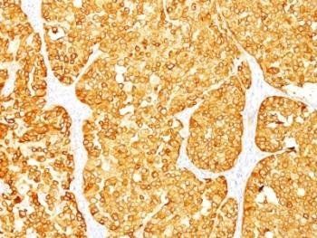
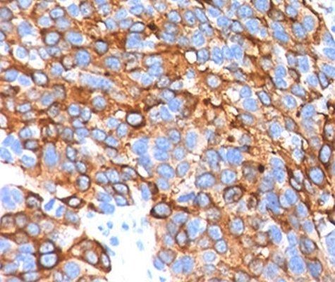
Immunohistochemical staining of Human melanoma using Melan-A Cocktail antibody
- Item 1 of 1
MART-1 / Melan-A Antibody Cocktail [orb248351]
FACS, IF, IHC-P, WB
Human, Mouse, Rat
Mouse
Monoclonal
Unconjugated
100 μg, 20 μg
Submit a review
Filter by Rating

- 5 stars

- 4 stars
- 3 stars
- 2 stars
- 1 stars