You have no items in your shopping cart.
Cart summary
Item 1 of 3
Item 1 of 3
MAP3K7 Antibody
Catalog Number: orb1246877

| Catalog Number | orb1246877 |
|---|---|
| Category | Antibodies |
| Description | MAP3K7 Antibody |
| Species/Host | Goat |
| Clonality | Polyclonal |
| Tested applications | ELISA, FC, WB |
| Predicted Reactivity | Bovine, Rat |
| Reactivity | Human, Mouse |
| Immunogen | The immunogen for this antibody is: C-AELDQDEKDQQNT |
| Concentration | 500 ug/mL |
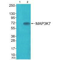
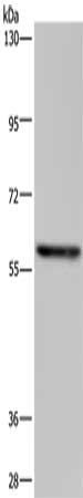
| Dilution range | Peptide ELISA: antibody detection limit dilution 1:8000.Western Blot:Approx 75kDa band observed in lysates of cell lines HeLa, Daudi and NIH3T3, and approx. 80kDa in lysates of cell line U937 (calculated MW of 67.2 according to Human NP_663304.1 and Mouse NP_033342.1). The observed molecular weight corresponds to different antibodies from other commercial sources. Recommended concentration: 1-3ug/ml. Primary incubation 1 hour at room temperature.Flow Cytometry: Flow cytometric analysis of HeLa cells. Recommended concentration: 10ug/ml. |
| Form/Appearance | Liquid |
| Conjugation | Unconjugated |
| Target | MAP3K7 |
| UniProt ID | O43318 |
| NCBI | NP_663304.1, NP_003179.1 |
| Storage | Aliquot and store at -20°C. Minimize freezing and thawing. |
| Buffer/Preservatives | Supplied at 0.5 mg/ml in Tris saline, 0.02% sodium azide, pH 7.3 with 0.5% bovine serum albumin. Aliquot and store at -20°C. Minimize freezing and thawing. |
| Alternative names | MAP3K7 , TAK1, mitogen-activated protein kinase ki Read more... |
| Note | For research use only |
| Application notes | Peptide ELISA: antibody detection limit dilution 1:8000.Western Blot:Approx 75kDa band observed in lysates of cell lines HeLa, Daudi and NIH3T3, and approx. 80kDa in lysates of cell line U937 (calculated MW of 67.2 according to Human NP_663304.1 and Mouse NP_033342.1). The observed molecular weight corresponds to different antibodies from other commercial sources. Recommended concentration: 1-3ug/ml. Primary incubation 1 hour at room temperature.Flow Cytometry: Flow cytometric analysis of HeLa cells. Recommended concentration: 10ug/ml. |
| Expiration Date | 12 months from date of receipt. |

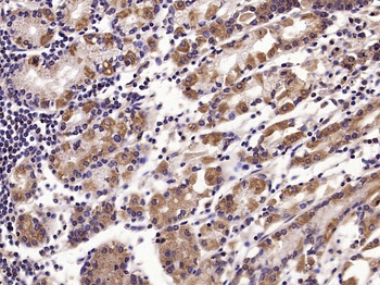
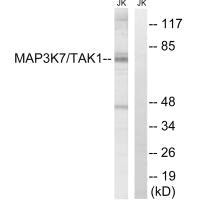
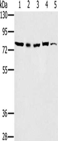
orb1246877 (1 ug/ml) staining of HeLa (A) and U937 (B) and (2 ug/ml) Daudi (C) cell lysate (35 ug protein in RIPA buffer). Detected by chemiluminescence.

orb1246877 (2 ug/ml) staining of NIH3T3 cell lysate (35 ug protein in RIPA buffer). Detected by chemiluminescence.

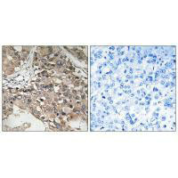
orb1246877 Flow cytometric analysis of paraformaldehyde fixed HeLa cells (blue line), permeabilized with 0.5% Triton. Primary incubation 1hr (10 ug/ml) followed by Alexa Fluor 488 secondary antibody (1 ug/ml). IgG control: Unimmunized goat IgG (black line).
- Item 1 of 5
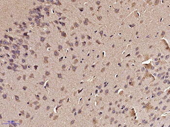
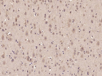
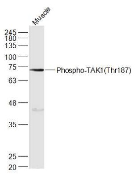
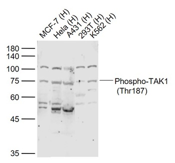
TAK1 (phospho-Thr187) antibody [orb7049]
FC, IHC-P, WB
Bovine, Equine, Gallus, Porcine, Rabbit
Human, Mouse, Rat
Rabbit
Polyclonal
Unconjugated
100 μl, 200 μl, 50 μl - Item 1 of 3
- Item 1 of 4
- Item 1 of 3
- Item 1 of 3
Submit a review
Filter by Rating

- 5 stars

- 4 stars
- 3 stars
- 2 stars
- 1 stars
