You have no items in your shopping cart.
Cart summary
Item 1 of 6
Item 1 of 6
LI Cadherin/CDH17 Antibody
Catalog Number: orb865373
| Catalog Number | orb865373 |
|---|---|
| Category | Antibodies |
| Description | LI Cadherin/CDH17 Antibody |
| Species/Host | Rabbit |
| Clonality | Polyclonal |
| Tested applications | ELISA, FC, ICC, IF, IHC |
| Reactivity | Human, Mouse, Rat |
| Isotype | Rabbit IgG |
| Immunogen | E.coli-derived human LI Cadherin/CDH17 recombinant protein (Position: E24-Q695). |
| Concentration | Adding 0.2 ml of distilled water will yield a concentration of 500 μg/ml. |
| Dilution range | Immunohistochemistry(Paraffin-embedded Section), 1-2 μg/ml, Human, Mouse, Rat Immunocytochemistry/Immunofluorescence, 5 μg/ml, Human Immunofluorescence, 5 μg/ml, Human Flow Cytometry(Fixed), 1-3 μg/1x106 cells, Human Direct ELISA, 0.1-0.5 μg/ml, Human |
| Form/Appearance | Lyophilized |
| Conjugation | Unconjugated |
| UniProt ID | Q12864 |
| Storage | At -20°C for one year from date of receipt. After reconstitution, at 4°C for one month. It can also be aliquotted and stored frozen at -20°C for six months. Avoid repeated freezing and thawing. |
| Note | For research use only |
| Application notes | Tested Species: In-house tested species with positive results. Other applications have not been tested. Optimal dilutions should be determined by end users. Adding 0.2 ml of distilled water will yield a concentration of 500 μg/ml. |
| Expiration Date | 12 months from date of receipt. |
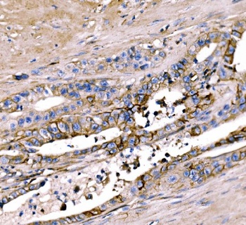
IHC analysis of LI Cadherin/CDH17 using anti-LI Cadherin/CDH17 antibody (orb865373). LI Cadherin/CDH17 was detected in a paraffin-embedded section of human appendicitis tissue. Heat mediated antigen retrieval was performed in EDTA buffer (pH 8.0, epitope retrieval solution). The tissue section was blocked with 10% goat serum. The tissue section was then incubated with 2 μg/ml rabbit anti-LI Cadherin/CDH17 Antibody (orb865373) overnight at 4°C. Biotinylated goat anti-rabbit IgG was used as secondary antibody and incubated for 30 minutes at 37°C. The tissue section was developed using Strepavidin-Biotin-Complex (SABC) (Catalog # orb90444) with DAB as the chromogen.
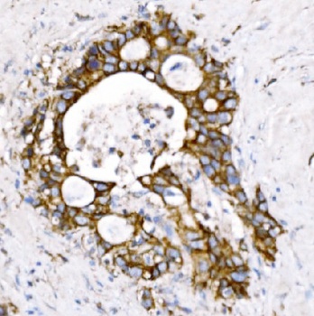
IHC analysis of LI Cadherin/CDH17 using anti-LI Cadherin/CDH17 antibody (orb865373). LI Cadherin/CDH17 was detected in a paraffin-embedded section of human gall bladder adenosquamous carcinoma tissue. Heat mediated antigen retrieval was performed in EDTA buffer (pH 8.0, epitope retrieval solution). The tissue section was blocked with 10% goat serum. The tissue section was then incubated with 2 μg/ml rabbit anti-LI Cadherin/CDH17 Antibody (orb865373) overnight at 4°C. Biotinylated goat anti-rabbit IgG was used as secondary antibody and incubated for 30 minutes at 37°C. The tissue section was developed using Strepavidin-Biotin-Complex (SABC) (Catalog # orb90444) with DAB as the chromogen.
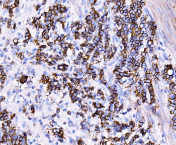
IHC analysis of LI Cadherin/CDH17 using anti-LI Cadherin/CDH17 antibody (orb865373). LI Cadherin/CDH17 was detected in a paraffin-embedded section of human stomach cancer tissue. Heat mediated antigen retrieval was performed in EDTA buffer (pH 8.0, epitope retrieval solution). The tissue section was blocked with 10% goat serum. The tissue section was then incubated with 2 μg/ml rabbit anti-LI Cadherin/CDH17 Antibody (orb865373) overnight at 4°C. Biotinylated goat anti-rabbit IgG was used as secondary antibody and incubated for 30 minutes at 37°C. The tissue section was developed using Strepavidin-Biotin-Complex (SABC) (Catalog # orb90444) with DAB as the chromogen.
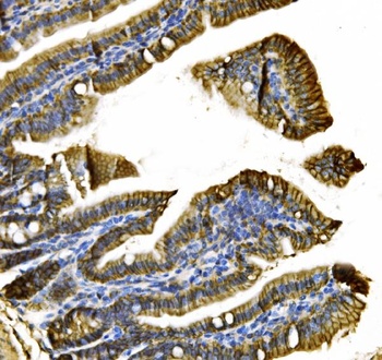
IHC analysis of LI Cadherin/CDH17 using anti-LI Cadherin/CDH17 antibody (orb865373). LI Cadherin/CDH17 was detected in a paraffin-embedded section of mouse colon tissue. Heat mediated antigen retrieval was performed in EDTA buffer (pH 8.0, epitope retrieval solution). The tissue section was blocked with 10% goat serum. The tissue section was then incubated with 2 μg/ml rabbit anti-LI Cadherin/CDH17 Antibody (orb865373) overnight at 4°C. Biotinylated goat anti-rabbit IgG was used as secondary antibody and incubated for 30 minutes at 37°C. The tissue section was developed using Strepavidin-Biotin-Complex (SABC) (Catalog # orb90444) with DAB as the chromogen.
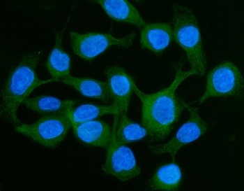
IF analysis of LI Cadherin/CDH17 using anti-LI Cadherin/CDH17 antibody (orb865373). LI Cadherin/CDH17 was detected in an immunocytochemical section of CACO-2 cells. Enzyme antigen retrieval was performed using IHC enzyme antigen retrieval reagent (orb90553) for 15 mins. The cells were blocked with 10% goat serum. And then incubated with 5 μg/mL rabbit anti-LI Cadherin/CDH17 Antibody (orb865373) overnight at 4°C. DyLight®488 Conjugated Goat Anti-Rabbit IgG was used as secondary antibody at 1:100 dilution and incubated for 30 minutes at 37°C. The section was counterstained with DAPI. Visualize using a fluorescence microscope and filter sets appropriate for the label used.
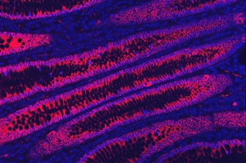
IF analysis of LI Cadherin/CDH17 using anti-LI Cadherin/CDH17 antibody (orb865373). LI Cadherin/CDH17 was detected in a paraffin-embedded section of human rectal cancer tissue. Heat mediated antigen retrieval was performed in EDTA buffer (pH 8.0, epitope retrieval solution). The tissue section was blocked with 10% goat serum. The tissue section was then incubated with 5 μg/mL rabbit anti-LI Cadherin/CDH17 Antibody (orb865373) overnight at 4°C. Biotin conjugated goat anti-rabbit IgG (orb1474935) was used as secondary antibody and incubated for 30 minutes at 37°C. The tissue section was developed using Cy3 Conjugated Avidin (orb27703). The section was counterstained with DAPI. Visualize using a fluorescence microscope and filter sets appropriate for the label used.

- Item 1 of 1

Submit a review
Filter by Rating

- 5 stars

- 4 stars
- 3 stars
- 2 stars
- 1 stars