You have no items in your shopping cart.
Cart summary
Item 1 of 3
Item 1 of 3
LCK (phospho-Y393) antibody
Catalog Number: orb214180
| Catalog Number | orb214180 |
|---|---|
| Category | Antibodies |
| Description | Rabbit polyclonal antibody to LCK |
| Species/Host | Rabbit |
| Clonality | Polyclonal |
| Tested applications | WB |
| Reactivity | Human, Mouse, Porcine, Rat, Sheep |
| Immunogen | KLH-conjugated synthetic phosphopeptide corresponding to residues surrounding Y393 of human LCK protein. The exact sequence is proprietary. |
| Dilution range | WB: 1-500-1-1000, IF/ICC: 1-100-1-500 |
| Form/Appearance | Liquid in 0.42% Potassium phosphate, 0.87% Sodium chloride, pH 7.3, 30% glycerol, and 0.01% sodium azide. |
| Conjugation | Unconjugated |
| Target | LCK |
| Entrez | 313050, 3932, 16818 |
| UniProt ID | P06240, Q01621, P06239 |
| Source | Rabbit |
| Storage | Shipped at 4°C. Upon delivery aliquot and store at -20°C for one year. Avoid freeze/thaw cycles. |
| Buffer/Preservatives | Liquid in 0.42% Potassium phosphate, 0.87% Sodium chloride, pH 7.3, 30% glycerol, and 0.01% sodium azide. |
| Alternative names | anti Tyrosine-protein kinase Lck antibody, anti Le Read more... |
| Note | For research use only |
| Expiration Date | 12 months from date of receipt. |
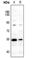
Western blot analysis of LCK (Phospho-Y393) expression in Myla2059 (A), HuT78 (B) whole cell lysates. (Predicted band size: 58 kD; Observed band size: 56 kD)
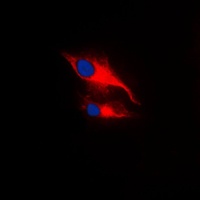
Immunofluorescent analysis of LCK (Phospho-Y393) staining in MCF7 cells. Formalin-fixed cells were permeabilized with 0.1% Triton X-100 in TBS for 5-10 minutes and blocked with 3% BSA-PBS for 30 minutes at room temperature. Cells were probed with the primary antibody in 3% BSA-PBS and incubated overnight at 4 °C in a humidified chamber. Cells were washed with PBST and incubated with a DyLight 594-conjugated secondary antibody (red) in PBS at room temperature in the dark. DAPI was used to stain the cell nuclei (blue).
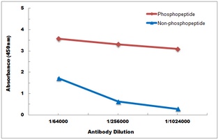
Direct ELISA antibody dose-response curve using Anti-LCK (Phospho-Y393) Antibody. Antigen (Phosphopeptide and non-phosphopeptide) concentration is 5 ug/ml. Goat Anti-Rabbit IgG (H&L) - HRP was used as the secondary antibody, and signal was developed by TMB substrate.

LCK (Phospho-Y393) antibody [orb571065]
ELISA, IF, WB
Human, Mouse, Rat
Rabbit
Polyclonal
Unconjugated
50 μg, 100 μg
Submit a review
Filter by Rating

- 5 stars

- 4 stars
- 3 stars
- 2 stars
- 1 stars